| Ileocecal fold | |
|---|---|
 Superior ileocecal fossa | |
 Inferior ileocecal fossa | |
| Details | |
| Identifiers | |
| Latin | plica ileocaecalis |
| TA98 | A10.1.02.421 |
| TA2 | 3788 |
| FMA | 16532 |
| Anatomical terminology | |
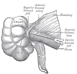
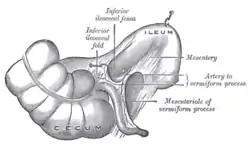
The ileocecal fold (or ileocaecal fold) is an anatomical structure of the human abdomen formed by a layer of peritoneum between the ileum and cecum. The upper border of the ileocecal fold is fixed to the ileum opposite its mesenteric attachment, and the lower border passes over the ileocecal junction to join the mesentery of the appendix (and sometimes the appendix itself as well). Behind the ileocecal fold is the inferior ileocecal fossa.[1]
The ileocecal fold is also called a ligament, veil, or bloodless fold of Treves (after English surgeon Sir Frederick Treves).[1] Despite the latter name, the ileocecal fold in fact often contains a vessel.[2]
Additional images
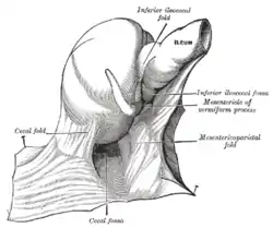
 The cecal fossa. The ileum and cecum are drawn backward and upward.
References
![]() This article incorporates text in the public domain from page 1160 of the 20th edition of Gray's Anatomy (1918)
This article incorporates text in the public domain from page 1160 of the 20th edition of Gray's Anatomy (1918)
- 1 2 Sir Frederick Treves Archived 2009-07-13 at the Wayback Machine at whonamedit.com
- ↑ Clinical Anatomy | Applied Anatomy for Students and Junior Doctors by Harold Ellis & Vishy Mahadevan - Thirteenth Edition (Page 88)
External links