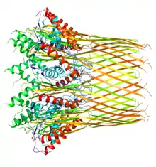
| Curli secretion channel | |||||||
|---|---|---|---|---|---|---|---|
 | |||||||
| Identifiers | |||||||
| Organism | |||||||
| Symbol | CsgG | ||||||
| Entrez | 945619 | ||||||
| PDB | 3X2R | ||||||
| RefSeq (mRNA) | NC_000913.3 | ||||||
| RefSeq (Prot) | NP_415555.1 | ||||||
| UniProt | P0AEA2 | ||||||
| Other data | |||||||
| Chromosome | Genomic: 1.1 - 1.1 Mb | ||||||
| |||||||
.pdf.jpg.webp)
The Curli protein is a type of amyloid fiber produced by certain strains of enterobacteria. They are extracellular fibers located on bacteria such as E. coli and Salmonella spp.[2] These fibers serve to promote cell community behavior through biofilm formation in the extracellular matrix. Amyloids are associated with several human neurodegenerative diseases such as Alzheimer's disease, Huntington's disease, Parkinson's disease, and prion diseases.[2][3] The study of curli may help to understand human diseases thought to arise from improper amyloid fiber formation.[2] The curli pili are generally assembled through the extracellular nucleation/precipitation pathway.[4]
Gene biosynthesis and regulation
A convenient way to identify genes important for curli production is by growing curliated bacteria on plates supplemented with Congo red diazo dye, which causes a red shift.[5] Curli fibers, and thus the curli protein, are coded by two divergently-transcribed operons in E. coli: the csgBAC and csgDEFG operons. In total, these operons encode at least seven proteins. The agfBA and agfDEFG operons, which have been identified in Salmonella spp., are homologous to the csgBAC and csgDEFG operons.[6][7] The csgBAC operon is responsible for the coding the three proteins CsgB, CsgA, and CsgC—all responsible for either the major subunit formation within the curli fiber or the inhibition of it.[7] The csgDEFG operon codes for proteins CsgD, CsgE, CsgF, and CsgG, and is responsible for the assembly, translocation, and regulation of the curli protein.[7]
CsgD is a positive transcriptional regulator of the csgBAC operon, but does not regulate its own expression. The csgBA operon encodes the major structural subunit of curli, CsgA, as well as the nucleator protein, CsgB. Further research still needs to be conducted in order to see what stimulates CsgD expression or activity, but some evidence suggests that its activation is caused by the phosphorylation of an aspartic acid residue of the N-terminal receiver domain. Since CsgD must be present for csgBA promoter activity, it is therefore suggested that regulators of CsgD expression also influence the expression of csgBA.[2]
Many environmental cues play a role in curli gene expression. It has long been believed that bacterial growth at a temperature below 30 °C promotes curli gene expression.[2] However, it is now known that curli expression is strain- and condition-specific. For example, mutations to the CsgD promoter can result in curli expression at 37 °C.[8] Additionally, when there is a lack of salt and nutrients such as nitrogen, phosphate or iron, curli gene expression is stimulated.[2]
Functions
Amyloids have been linked to illnesses such as Alzheimer's, Parkinson's, Huntington's, lupus, among many others.[7][9] The curli protein is considered a PAMP (Pathogen-associated molecular pattern); its β-sheet structure is recognized in the innate immune system and activates the TLR2 (Toll-Like Receptor 2).[7] This then causes a downstream response by producing a pro-inflammatory response where pro-inflammatory cytokines and chemokines are recruited to initiate the inflammation response.[7]
Curli, however, has additional functions (thus being coined a "functional amyloid"[9]) including being a major component in the biofilm generated by gram-negative bacteria such as E. coli and Salmonella spp.[6][7][9] These biofilms allow gram-negative bacteria to better colonize in a given environment, protecting them from oxidative stress and dehydration.[6][7] These biofilms, however, call for much concern. Since these biofilms allow for the bacteria to survive chemical and physical stressors within their environment, not only do they make patients more susceptible to infection when using shared appliances, but curli and other biofilms have been shown to reduce the infected individuals' immune response and antimicrobials.[6] Curli proteins and biofilms alike are very resistant to chemical stressors to a point where stronger pretreatment is required in order for curli to degrade or dissolve in sodium dodecyl sulphate (SDS).[6]
Curli fibers that are created are involved in many different types and functions in the cell such as for cell aggregation, act as adhesive for different proteins to the cell's surface, used in biofilm formation of certain bacteria and contain chemicals for quorum sensing, as well as plays an important role in inflammatory response in individuals.[10] Curli formation must also be heavily regulated as its accumulation leads to amyloid like protein aggregations in the organism which leads to destruction of pathway and interferes with cell signaling.[11]
Structure
The curli protein's main components (subunits) consist of the CsgA and CsgB protein.
CsgA
CsgA is the major subunit of the curli protein and weighs approximately 13.1 kilodalton. This protein consists of three domains which have a tendency to aggregate and form amyloid fibrils: a single peptide, a 22-amino acid N-terminal sequence (used for secretion), and an amyloid core domain at the C-terminal sequence.[6][9] The amyloid core domain is composed of 5 repeating (yet not exact) sequences revolving around the sequence: Ser-X5-Gln-X-Gly-X-Gly-Asn-X-Ala-X3-Gln.[9] This repeating sequence is the characteristic subunit that forms its aggregatable β-sheet.[9]
CsgB
CsgB, also known as the minor subunit, is required for the nucleation and organization of CsgA into a curli fiber on cell surfaces.[7] CsgB has a very similar repeating sequence to that of CsgA with the exception that one of the 5 repeating sequences contains additional amino acids: Lys133, Arg140, Arg14, and Arg151.[6] This change in the final subunit (known as the R5 subunit) is required. Without the presence of the R5 subunit, or the changes within the subunit, CsgA is secreted from the cell in an unpolymerized form and cannot properly form on cell surfaces.[6][9]
CsgC
The CsgC subunit was only recently discovered to prevent the aggregation and polymerization of the CsgA protein. Without it, there is a chance for amyloid fibril formation and even cell death.[7] Multiple experiments isolating CsgC away from the CsgA and CsgB subunit caused CsgA to aggregate into fibrils, which may possibly lead to downstream effects as seen in illnesses such as Alzheimer's.[6] The molar ratio required for CsgC to inhibit CsgA is 1:500, meaning only 1 CsgC protein is required to inhibit 500 CsgA proteins from forming amyloid fibril structures.[6][12] It is hypothesized that CsgC is therefore considered a chaperone since it prevents further CsgA nucleation and allows CsgA to form into its proper structure instead of aggregating.[6]
References
- ↑ Praveschotinunt P, Duraj-Thatte AM, Gelfat I, Bahl F, Chou DB, Joshi NS (December 2019). "Engineered E. coli Nissle 1917 for the delivery of matrix-tethered therapeutic domains to the gut". Nature Communications. 10 (1): 5580. Bibcode:2019NatCo..10.5580P. doi:10.1038/s41467-019-13336-6. PMC 6898321. PMID 31811125.
- 1 2 3 4 5 6 Barnhart MM, Chapman MR (2006). "Curli biogenesis and function". Annual Review of Microbiology. 60: 131–147. doi:10.1146/annurev.micro.60.080805.142106. PMC 2838481. PMID 16704339.
- ↑ Chapman MR, Robinson LS, Pinkner JS, Roth R, Heuser J, Hammar M, et al. (February 2002). "Role of Escherichia coli curli operons in directing amyloid fiber formation". Science. 295 (5556): 851–855. Bibcode:2002Sci...295..851C. doi:10.1126/science.1067484. PMC 2838482. PMID 11823641.
- ↑ Hammar M, Bian Z, Normark S (June 1996). "Nucleator-dependent intercellular assembly of adhesive curli organelles in Escherichia coli". Proceedings of the National Academy of Sciences of the United States of America. 93 (13): 6562–6566. Bibcode:1996PNAS...93.6562H. doi:10.1073/pnas.93.13.6562. PMC 39064. PMID 8692856.
- ↑ Collinson SK, Doig PC, Doran JL, Clouthier S, Trust TJ, Kay WW (January 1993). "Thin, aggregative fimbriae mediate binding of Salmonella enteritidis to fibronectin". Journal of Bacteriology. 175 (1): 12–18. doi:10.1128/jb.175.1.12-18.1993. PMC 196092. PMID 8093237.
- 1 2 3 4 5 6 7 8 9 10 11 Van Gerven N, Klein RD, Hultgren SJ, Remaut H (November 2015). "Bacterial amyloid formation: structural insights into curli biogensis". Trends in Microbiology. 23 (11): 693–706. doi:10.1016/j.tim.2015.07.010. PMC 4636965. PMID 26439293.
- 1 2 3 4 5 6 7 8 9 10 Tursi SA, Tükel Ç (December 2018). "Curli-Containing Enteric Biofilms Inside and Out: Matrix Composition, Immune Recognition, and Disease Implications". Microbiology and Molecular Biology Reviews. 82 (4): e00028–18, /mmbr/82/4/e00028–18.atom. doi:10.1128/MMBR.00028-18. PMC 6298610. PMID 30305312.
- ↑ Uhlich GA, Keen JE, Elder RO (May 2001). "Mutations in the csgD promoter associated with variations in curli expression in certain strains of Escherichia coli O157:H7". Applied and Environmental Microbiology. 67 (5): 2367–2370. Bibcode:2001ApEnM..67.2367U. doi:10.1128/AEM.67.5.2367-2370.2001. PMC 92880. PMID 11319125.
- 1 2 3 4 5 6 7 Evans ML, Chapman MR (August 2014). "Curli biogenesis: order out of disorder". Biochimica et Biophysica Acta (BBA) - Molecular Cell Research. 1843 (8): 1551–1558. doi:10.1016/j.bbamcr.2013.09.010. PMC 4243835. PMID 24080089.
- ↑ Botyanszki, Zsofia; Tay, Pei Kun R.; Nguyen, Peter Q.; Nussbaumer, Martin G.; Joshi, Neel S. (October 2015). "Engineered catalytic biofilms: Site-specific enzyme immobilization onto E. coli curli nanofibers: Catalytic Biofilms Using Engineered Curli Fibers". Biotechnology and Bioengineering. 112 (10): 2016–2024. doi:10.1002/bit.25638. PMID 25950512. S2CID 9350467.
- ↑ Evans, Margery L.; Chapman, Matthew R. (August 2014). "Curli biogenesis: order out of disorder". Biochimica et Biophysica Acta (BBA) - Molecular Cell Research. 1843 (8): 1551–1558. doi:10.1016/j.bbamcr.2013.09.010. ISSN 0006-3002. PMC 4243835. PMID 24080089.
- ↑ Evans ML, Chorell E, Taylor JD, Åden J, Götheson A, Li F, et al. (February 2015). "The bacterial curli system possesses a potent and selective inhibitor of amyloid formation". Molecular Cell. 57 (3): 445–455. doi:10.1016/j.molcel.2014.12.025. PMC 4320674. PMID 25620560.
Further reading
- Dema B, Charles N (January 2016). "Autoantibodies in SLE: Specificities, Isotypes and Receptors". Antibodies. 5 (1): 2. doi:10.3390/antib5010002. PMC 6698872. PMID 31557984.
- Gallo PM, Rapsinski GJ, Wilson RP, Oppong GO, Sriram U, Goulian M, et al. (June 2015). "Amyloid-DNA Composites of Bacterial Biofilms Stimulate Autoimmunity". Immunity. 42 (6): 1171–1184. doi:10.1016/j.immuni.2015.06.002. PMC 4500125. PMID 26084027.
- Kono DH, Baccala R, Theofilopoulos AN (December 2013). "TLRs and interferons: a central paradigm in autoimmunity". Current Opinion in Immunology. 25 (6): 720–727. doi:10.1016/j.coi.2013.10.006. PMC 4309276. PMID 24246388.
- Tursi SA, Lee EY, Medeiros NJ, Lee MH, Nicastro LK, Buttaro B, et al. (April 2017). "Bacterial amyloid curli acts as a carrier for DNA to elicit an autoimmune response via TLR2 and TLR9". PLOS Pathogens. 13 (4): e1006315. doi:10.1371/journal.ppat.1006315. PMC 5406031. PMID 28410407.