Case study
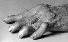
- The patient is a 57 year old female. Retired nurse was diagnosed with RA (Rheumatoid Arthritis) at the age of 25. At that time, she was working as a nurse in a clinic of a primary school. They started with her the different medicines used with RA patients. During the first few years she wasn't regular on her medicine. After 10 years of diagnosis she started to notice her fingers to look strange and the doctor told her that she is developing a well-known deformity, that is Swan neck deformity. She was then referred to the orthotic department, were she was given a special made orthosis for her deformity. At first she was given a custom-made finger rings that she used for few days. However, due to pain and discomfort, she stopped wearing them few days later. Then she was given a prefabricated readymade finger rings. She used them for few weeks while working, but with these also she was feeling discomfort and pain while wearing them during work so she stopped using them.
Evidence
- Swan neck deformity (SND) of the fingers is a well-known deformity seen mostly in Rheumatoid Arthritis’ patient and has been recognized clinically for perhaps 300 years ago (Dreyfus & Thomas, 1983). The term SND has only been used since 1957 introduced by Laine et. al. (Dreyfus & Thomas, 1983). It is also known as the deformity causing the hyperextension of the proximal interphalangeal joint with reciprocal flexion of metacarpophalangeal and distal interphalangeal joints (Heywood, 1979). There are various types of swan neck deformity. Although the pathological process may be the same, the swan neck deformities character may differ not only from patient to patient, but also from finger to finger in the same hand (Welsh & Hastings, 1977). Some reasons that can cause swan neck deformity: EHLERS-DANLOS Syndrome (Ercocen, Yenidunya, Yilmaz, & Ozbek, 1997), Rheumatoid Arthritis (Knight, 2006), Dislocation of a finger by AGEE Technique – as a complication of treating a fracture (Rawes & Oni, 1995), Contracture post injury to the volar aspect of the proximal interphalangeal flexion crease (Chinchalkar, Lanting & Ross, 2010), and Congenital swan neck deformity secondary to laxity of the palmar plate of the proximal interphalangeal joint (Ozturka, Zorb, Sengezera & Isik, 2005).
- The SND has a great impact on patients' functional ability to perform their daily tasks. It is important to gain a knowledgeable understanding of this deformity by understanding the structural and dynamic factors that constitute the normal muscle and tendon balance of the digits (Dreyfus & Thomas, 1983). They consist of 3 sets of muscles and tendons responsible for the equilibrium of the digits, and they are; the extrinsic extensor group, the extrinsic flexor group, and the intrinsic muscles (Dreyfus & Thomas, 1983). The extrinsic extensor group of muscles includes the extensor digitorum communis, extensor digiti minimi and the extensor indicis. These muscles originate in the dorsal forearm and insert via the common extensor tendons into the extensor hood and onto the dorsal surface of each digit. The sites of insertion are multiple and include the dorsal base of the proximal and middle phalanges as well as the distal phalanx via the lateral extensor tendons. The nerve supply is a branch of the radial nerve. The extensor muscles act via the extensor mechanism to extend the digits at the metacarpophalangeal (MCP) joint. In concert with the intrinsic muscles, they extend the proximal interphalangeal (PIP) and the distal interphalangeal DIP joints as well. The extrinsic flexor group includes the flexor digitorum profundus and superficialis (the deep and superficial flexors), originating in the volar forearm and inserting respectively into the bases of the distal and middle phalanges. The extrinsic flexors, supplied by branches of the median and ulnar nerves, flex the fingers at the PIP and DIP joints. Efficient MCP flexion requires the synergistic action of the intrinsic muscles.
- The intrinsic muscles (IM) include the four lumbricals and the seven volar and dorsal interossei. The lumbricals arise from the flexor tendons and insert into the lateral extensor bands. The interossei arise between the metacarpals and have various insertions in the MCP joint capsule and proximal phalangeal base, the extensor hood, and the medial and lateral extensor bands. The usual nerve supply of these muscles is the ulnar nerve with the exception of the first and second lumbricals, which are supplied by the median nerve. The intrinsic muscles act as a functional unit with two major effects: flexion of the MCP joints and extension of the PlP and DIP joints. By virtue of insertions into the base of the proximal phalanx volar to the axis of the MCP joint, the intrinsic muscles flex the MCP. The tendons then cross dorsally and join the extensor apparatus over the PIP and thereby act to extend the PIP and DIP (Dreyfus & Thomas, 1983).
Orthotic treatment options
- The conventional technique used in the treatment of the different kinds of the swan neck deformity is Hand Orthosis. There are many types of these orthotic devices that result in different outcomes. Patients with swan neck deformity experience a lot of problems; the main ones are pain and limitation of functional daily activities. Either fixed or flexible, the swan neck deformity, may limit the ability to actively flex the PIP joint, with negative consequences for performing a pinch grip and grasping larger objects with one hand (Van der Giesen, Nelissen, Van Lankveld, Kremers-Selten, Peeters, Stern, Cessie & Vlieland, 2010). Studies on the effectiveness of the different types of splints used for swan neck deformity are little. There are 3 to 4 known types of the splints used for swan neck deformities. Silver ring splints, custom-made thermoplastic splints and prefabricated thermoplastic splints (Van der Giesen, Nelissen, Van Lankveld, Kremers-Selten, Peeters, Stern, Cessie & Vlieland, 2010). The main aim of using these splints is to prevent the deformity from becoming worse and to give the patient more functional use in his/her daily activities. All these splints are based on the same principle of using a three - point force around the PIP joint to prevent hyperextension at the PIP joint and subsequent DIP joint flexion- i.e. swan neck deformity (Adams, Hammond, Burridge & Cooper, 2005). Literature showed that several studies have been carried out to compare the effectiveness of the different types of the available splints. All these studies have shown the effectiveness of several types of the orthotic devices used by therapists in treating swan neck deformity.
- Knipping & Schegget (2000) compared the use and effects of custom- made versus prefabricated splints for swan neck deformities in Rheumatoid Arthritis patients on the variables cosmesis, comfort, functional use, and wearing time per 24 hours and effects on stability, grip strength and mobility. It was carried out on 18 patients 9 in each group. Each group used one of the splints for 3 months and another 3 months used the other splint. They concluded that the prefabricated splints are tolerated better and more acceptable in terms of wear than the custom- made splints. Zijlstra, Heijnsdijk-Rouwenhos, & Rasker (2004) studied the effect of Silver Ring Splints on hand function in patients with RA. They concluded that the Silver Ring splint could improve dexterity in patients with RA. In 2009, Van der Giesen, Nelissen, Van Lankveld, Kremers-Selten, Peeters, Stern, Cessie & Vlieland done a study on the effectiveness of silver ring splints with the commercial prefabricated thermoplastic splints in RA patients. They came to a conclusion that with RA patients with a mobile swan neck deformity the 2 kinds of splints are equally effective and acceptable. In 2010, Van der Giesen, Nelissen, Van Lankveld, Kremers-Selten, Peeters, Stern, Cessie & Vlieland done a qualitative study on patients' perspectives on hand function problems and finger splints to identify hand function problems and the reasons for choosing a specific finger splint in patients with RA and swan neck deformities to find that these 2 kinds of splints have the same effectiveness. In 2012, Elui, Rodrigues, Purquerio, Fortula & Fonseca presented the development of a splint with the original concept of blocking hyperextension of PIP joint in order to increase the functionality of the hand in carrying out daily activities. This splint defers from the old ones that the 2 separate parts joined with an axis of rotation allowing more function in the fingers. The study showed that this model has a satisfactory engagement with the finger, corrected the deformity and minimized the functional difficulties experienced by the patient daily.
Comparison of Swan Neck deformity treatment options
- Despite the fact that swan neck deformity is usually treated with Hand Orthoses, a lot of different surgical procedures have been carried out in order to correct the swan neck deformity to increase functional daily activities levels. Cosmetic reasons have also been related to several surgical interventions. There are many surgical ways of treating SND, some of them are; distal joint infusion, dermadesis, flexor tendon tenodesis, retincular ligament reconstruction, intrinsic release, lateral band mobilization, skin release, arthodesis of the PIP joint, and arthroplasty with silicone rubber implants (Gainor & Hummel, 1985). A correction of the problem can also be by re-routing a lateral band of the extensor mechanism to prevent extension of the proximal PIP joint (Sirotakova, Figus, Jarrett, Mishra & Elliot, 2008). With many surgical ways of treatments to the deformity, Dreyfus & Thomas (1983) suggests that the key to the selection of the appropriate surgical treatment depends exclusively on identification of the pathogenetic mechanism.
References
- Adams, J., Hammond, A., Burridge, J., & Cooper, C. (2005). Static orthoses in the prevention of hand dysfunction in rheumatoid arthritis: A review of the literature. Musculoskeletal Care, 3(2), 85-101
- Chinchalkar, S. J., Lanting, B. A., & Ross, D. (2010. Oct-Dec). Swan Neck Deformity after Distal Interphalangeal Joint Flexion Contractures: A Biomechanical Analysis. Journal of Hand Therapy, 420-425
- Dreyfus, J. N., & Thomas J. S. (1983, November). Pathogenesis and Differential Diagnosis Swan-Neck Deformity. Seminars in Arthritis and Rheumarism, 13(2), 200-211
- Elui, V. M., Rodrigues, M. C., Purquerio, B. M., Fortulan, C. A., & Fonseca, M. R. (2012, Oct-Dec). Swan Neck Deformity Orthosis Development: Aoa (Articulated Ring Orthosis). Journal of Hand Therapy, 25(4), e9-e10
- Ercocen, A. R., Yenidunya, M. O., Yilmaz, S., & Ozbek, M. R. (1997). Dynamic Swan Neck Deformity in a Patient with Ehlers-Danlos Syndrome. Journal of Hand Surgery, 22B(1), 128-130
- Gainor, B. J., & Hummel, G. L. (1985, May). Correction of rheumatoid swan-neck deformity by lateral band mobilization. The Journal Of Hand Surgery, 10A(3), 370-376
- Heywood, A. W. B. (1979). The Pathogenesis of the Rheumatoid Swan Neck Deformity. The Hand, 11(2), 176-183
- Knight, S. L. (2006). Assessment and management of swan neck deformity. International Congress Series, 154–157
- Knipping, A., & Schegget, M. t. (2000). A Study Comparing Use and Effects of Custom-Made versus Prefabricated Splints for Swan Neck Deformity in Patients with Rheumatoid Arthritis. The British Journal of Hand Therapy, 5, 101-107
- Ozturka, S., Zorb, F., Sengezera, M., & Isik, S. (2005). Correction of bilateral congenital swan-neck deformity by use of Mitek mini anchor: a new technique. British Journal of Plastic Surgery, 58, 822–825
- Rawes, M. I., & Oni O. O. A. (1995). Swan-Neck Deformity as a Complication of the Agee Technique. Journal of Hand Surgery, 20B(2), 255-257
- Sirotakova, M., Figus, A., Jarrett, P., Mishra, A., & Elliot, D. (2008). Correction of Swan Neck Deformity in Rheumatoid Arthritis Using a New Lateral Extensor Band Technique. The Journal of Hand Surgery, 33(6), 712–716
- Van der Giesen, F. J., Nelissen, R. G. H. H., Van Lankveld W. J., Kremers-Selten, C., Peeters, A. J., Stern, E. B., Cessie, S. l., & Vlieland, V. T. P. M. (2009, August 15). Effectiveness of Two Finger Splints for Swan Neck Deformity in Patients With Rheumatoid Arthritis: A Randomized, Crossover Trial. Arthritis & Rheumatism, 61(8), 1025–1031
- Van der Giesen, F. J., Nelissen, R. G. H. H., Van Lankveld W. J., Kremers-Selten, C., Peeters, A. J., Stern, E. B., Cessie, S. l., & Vlieland, V. T. P. M. (2010, May 11). Swan Neck Deformities in Rheumatoid Arthritis: A Qualitative Study on the Patients’ Perspectives on Hand Function Problems and Finger Splints. Finger Splints for Swan Neck Deformities in RA, 179-188
- Welsh, R. P., & Hastings, D. E. (1977). Swan Neck Deformity In Rheumatoid Arthritis Of The Hand. The Hand, 9(2), 109-116
- Zijlstra, T. R., Heijnsdijk-Rouwenhost, L., & Rasker, J. J. (2004, December 15). Silver Ring Splints Improve Dexterity in Patients With Rheumatoid Arthritis. Arthritis & Rheumatism, 51(6), 947-951
Search Strategy
Search undertaken for this assignment included various synonyms to help find a more abroad search. Databases searched were Midline, Cochrane and Google Scholar. Search terms "Swan Neck Deformity", "ortho*", "orthos*", "brace", and "splint" were used. Combinations of truncations and Boolean operators were also used for a wider range of results. Some limits such as English language and Humans were also used to specify the search. The results found a combination of recent and old studies, but only the relevant and most appropriate information addressing the topic was chosen.
Functional Aims and Goals
- The main aim of a finger splint in the treatment of Swan neck deformity is to prevent structural deterioration of the joint. Reduction of pain and swelling, increase ROM, and limiting hyperextension of the joint are also goals of the splint by the idea of resting the joints affected in position and basically limiting joint movement. Recent studies showed that the most effective orthosis used in patients with swan neck deformity nowadays are the articulated silver ring orthosis. The model has a satisfactory engagement with the finger, correcting the deformity and has synchronicity between finger movement and orthotic device, which minimises functional difficulties. However, due to lack of materials, we aimed for the same outcome but with a custom made finger splint using thermoplastic material. The custom made thermoplastic splint covered the palmar aspect of the affected finger ending at the MCP with 2 straps attached on the dorsal aspect. The permanent side of each strap was placed laterally to the index finger, in order to allow more space for adjustment medially and to make less of the material used laterally (between the index and the adjacent finger). Although it is a lot less functional for the patient, however, it will still achieve the same outcome using the 3 point force principle.
Design
- Material
- • The material used for this orthotic treatment is Low Temperature Thermoplastic.
- Trimlines and attachments
- • LTT finger splint covering palmer side of affected finger ending at MCP joint
- • Trimlines covering half of the circumference of the finger
- • Permanent side of straps placed lateral to index finger (allow more space for adjustment medially)
- • 2 straps attached on the dorsal side of the splint as seen on the initial design (Fig 1)
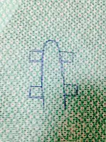 Fig 1 |
- Forces Diagram
- • Forces applied by the splint are demonstrated in sagittal view using the 3 force principle (Fig 10)
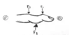 Sagittal View of the three force principle applied on the finger (Figure 10) |
Manufacturing process
- Tools required
- • Chair & table
- • Pen, ruler, scissors & goniometer
- • Plain cloth or paper to make pattern
- • LTT material
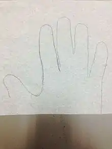 Fig 2
Fig 2
- • LTT material
- • Sticky back Velcro loop & Velcro hook
- • Electric fry pan
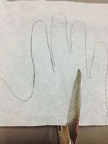 Fig 3
Fig 3
- • Electric fry pan
- • Heat gun
- Process
- • Position the patient sitting comfortably with his wrist and on a table
- • Have the patient’s hands placed on a piece of paper and draw around it to get the correct measurements (Fig 2)
- • Cut the drawn on piece of paper using a scissors (Fig 3)
- • Place the cut piece paper onto your LTT piece (Fig 4)
- • Trace the piece of paper onto the LTT piece and also marks of the joint places on it (Fig 5)
- • Cut around the trimlines you have drawn onto the LTT piece (Fig 6)
- • Dip the LTT piece into the hot water in the electric fry pan (Fig 7)
- • Remove LTT piece from water and let it slightly dry before applying it to patient
- • Apply the LTT piece on the patient with his palmar surface with hands placed in a supine position (eliminating gravity)
- • Mould the LTT piece on the patient’s finger held in required position and let it set
- • Draw on the LTT piece where you would want to get the any excess material to have more accurate trimlines for function (Fig 8)
- • Remove the piece and remove excess material that is not aligned with your trimlines
- • Trim lines are neaten by submerging the LTT piece back into the warm water and trimming the edges whiles it still hot
- • Fit the device on the patient again and see if what you have fixed has been corrected incase of you need any more modifications to the trim lines
- • Use acetone to remove any marks of your trim lines on the device
- • Applying the Velcro hooks on the dorsum side of the device just below the DIP & PIP joints
- • The two Velcro Straps are cut according to the needed length making the edges rounded to be neat
- • Heat the end of the Velcro loop strap using a heat gun and permanently bond them together (Fig 9)
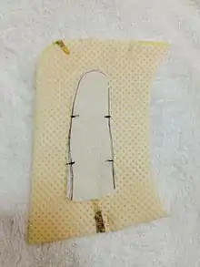 Fig 4 | 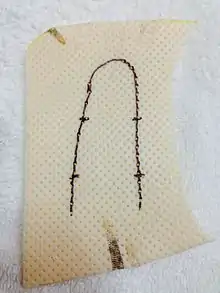 Fig 5 | 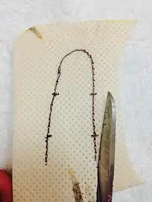 Fig 6 |  Fig 7 | 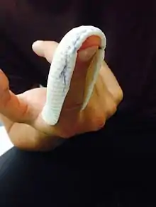 Fig 8 |
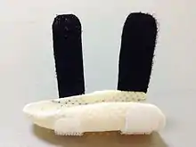 Fig 9 |
Critique of fit
- This is a splint made to correct the deformity of swan neck deformity characterized by hyperextension of PIP and flexion of DIP. The aim of the device is to correct this deformity by preventing the hyperextension and the flexion. The device made seen on (Fig 12) was fit and the aim of the device was reached. The trim lines were not neat and the drawing lines were obvious. Although acetone was used to clear them, trim lines that were drawn earlier in the process of making the device are still appearing on the device (Fig 13). The ROM was achieved at the MCP joint (Fig 14), which was one of the goals when making the splint, thus allowing the fingers to function as normal as possible during daily activities as seen on the figures (15 & 16). Concerning the straps, straps were slightly wide than needed in relation to the small size of the splint applying more force to achieve the required goals. Also, a softer material on the lateral side of the finger could have been used on the straps to prevent the discomfort a patient may feel from the straps between is fingers. Recent studies suggest that more beneficial splints used in such cases are the custom made articulated 8 oval rings splints made of silver (Elui et al, 2012). These types of splints allow more functional hands during daily activities while preventing the hyperextension of the PIP and flexion of DIP joints. This is by allowing flexion of the PIP and DIP joints during the activities and minimizing functional difficulties. For example, when a patient is holding a water bottle as seen on figure 15, a flexion of the PIP and DIP joints will be more functional for a better grip, this can only be achieved by using the recent articulated ring orthoses. Preventing the hyperextension of the PIP and flexion of DIP are still maintained, as they are the main goals of the splint. However, due to the lack of materials a regular splint preventing hyperextension of PIP and flexion of DIP was made using LTT material.
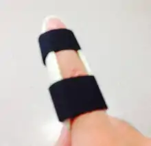 Fig 12 | 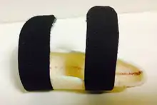 Fig 13 | 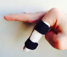 Fig 14 | 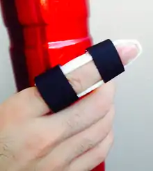 Fig 15 | 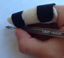 Fig 16 |
Outcome measures
- The aim of any orthotic device in treating the swan neck deformity is to correct the deformity and to minimize the functional difficulties, e.g. the 8 oval rings device. In order to see whether or not the orthotic device has reached the aims and goals, an outcome measure should be selected to measure the correction of the deformity and level of minimizing the functional difficulties. In this case, the quick Disability of Arm, Shoulder and Hand (DASH) questionnaire was used to measure the outcome of the swan neck deformity. The quick DASH measure was chosen because it consists of 11 items that measure the physical function and symptoms in people with any musculoskeletal disorders of the upper limb. Studies showed that the quick DASH is a comparable instrument and a more efficient version of the DASH outcome measure (Beaton, Wright & Katz. 2005). Although there is a full version of the DASH questionnaire that consists of 30 items to measure a similar outcome, the shorter version of the questionnaire (QuickDASH) was used as it was found to be more precise, attractive and a sensible option for a patient with swan neck deformity. Although there is a number of instruments that are used to assess the function of the hand and wrist joints, the quick DASH measure is believed to be the most widely used instrument for its reliability and validity for both clinical and research purposes (Hoang-Kim, Pegreffi, Moroni & Ladd, 2011).The 11 items in the quick DASH questionnaire were precise on measuring the activity levels of the related joints during daily activities. At least 10 of the 11 items have to be completed in order to have a calculated accurate score. Not only are these items repeatable, they are also easy to implement, short, simple and precise. Therefore, the quick DASH questionnaire has achieved what had hoped to be measured for patients with swan neck deformities. After administering the quick DASH questionnaire on the patient, before (figure 17) and after (figure 18) using the orthotic device, the calculated scores showed not much of a difference between the results (59.1 before and 50.0 after), showing that the patient did not benefit from the orthotic device as much as it was hoped.
 Fig 17 |  Fig 18 |
References
- Beaton, D. E., Wright, J. G., & Katz, J. N. (2005). Development of the QuickDASH: comparison of three item-reduction approaches. The Journal of bone and joint surgery, 87(5), 1038-1046
- Hoang-Kim, A., Pegreffi, F., Moroni, A., & Ladd, A. (2011). Measuring wrist and hand function: Common scales and checklists. Injury, 42(3), 253-258
- The DASH Outcome Measure (2014). Disabilities of the Arm, Shoulder and Hand. Retrieved from http://www.dash.iwh.on.ca
Referral
29th of May 2014
Dr John,
Melbourne Occupational Therapy Clinic 123 Bourke Street
Melbourne, VIC 300
Thanks for seeing Sarah Ronaldo. Sarah is a 57 yrs old female diagnosed with Swan neck deformity. She was also diagnosed with RA at the age of 25. She has been on painkillers and steroids since then. A costume made orthotic brace has been made so far in terms of Sarah’s management. Sarah has difficulties doing her daily activities like opening a jar and buttoning her shirt. Sarah has been very cooperative with her treatment. Please see what you could do in providing daily activities that would improve her range of motion.
Yours sincerely,