Describe your case study
The patient is a seventeen-year-old female who has experienced an accident whilst riding a horse two days ago. As she fell of her horse she landed on her right hand incorrectly, twisting her wrist. She experiences pain when rotating her forearm and has an excessive prominence of the ulnar head. Medical imaging shows a dorsal displacement ulna head and a partial tear of the triangular fibrocartilaginous complex (TFCC). The patient was diagnosed with an isolated dorsal distal radioulnar joint (DRUJ) dislocation. She is currently not using any medication and is commencing her final year of high-school and wishes to recover as soon as possible in order to complete studies efficiently.
Written information
Evidence
ANATOMY
The DRUJ is a complex articulation of the forearm involving the distal ends of the radius and ulna. The ulna is fixed in position as the radius rotates around (Sammer & Chung, 2012, p. 199). Movements of the DRUJ include supination; produced by biceps brachii and supinator and pronation; produced by pronator teres and pronator quadratus (Moore, Dalley & Agur, 2013, p. 806). This synovial joint consists of bony articulations and soft tissue stabilisers. The joint articulations are the sigmoid notch of the radius and the ulnar head. Due to the shape of the sigmoid notch, the joint lacks congruency; therefore soft tissue stabilisers maintain joint integrity (Mulford & Axelrod, 2010, p. 155). The TFCC is the primary stabiliser of the DRUJ, which originates from the radial sigmoid notch and inserts into the ulnar styloid. The TFCC complex consists of articular discs, dorsal and volar radioulnar ligaments, meniscal homologue, ulnar collateral ligament and the extensor carpi ulnaris subsheath (Micucci & Schmidt, 2007, p. 118). DRUJ injuries can be isolated or associated with fractures.
PATHOLOGY
DRUJ dislocations are commonly associated with fractures of the radius or ulna. Intra-articular fractures will disrupt the articulation of the joint. Extra-articular fractures may result in displacement, shortening, angulation or translation of the radius, which can cause DRUJ instability (Sammer & Chung, 2012, p. 199). Although dislocations of the DRUJ often occur in conjunction with fractures, there are also cases of isolated dislocations (Wassink, Lisowski & Schutte, 2009, p. 141; O'Brien & Thurn, 2013, p. 287-288). Common mechanisms of injury for DRUJ dislocations include; forced pronation/ supination, direct force to the ulna/ radius or fall on an outstretched hand (Wassink, Lisowski & Schutte, 2009, p. 143; Mulford & Axelrod, 2010, p. 156). DRUJ dislocations may involve damage to the primary stabiliser. Injury to the TFCC increases in severity in relation to force (Melone Jr & Nathan, 1992, p. 495-496), this will affect the stability of the joint. DRUJ dislocations can be stable or unstable, reducible or irreducible. Stable dislocations only have instability in particular positions and may have strains to the TFCC (Mulford & Axelrod, 2010, p. 156-157). Unstable dislocations have instability in all positions and are commonly associated with rupture or avulsion of the TFCC from ulnar styloid (Thomas & Sreekanth, 2012, p. 497). Irreducible DRUJ dislocations have underlying factors, such as soft tissues or bone, that block reduction (Mulford & Axelrod, 2010, p. 158). Untreated dislocations can lead to chronic instability or reduced range of motion. Depending on severity of the dislocation and other components involved, bone or soft tissue, appropriate treatment plan would be prescribed.
SPECIFIC TO CASE STUDY
Isolated dislocations of the DRUJ can be classified as volar, or dorsal (O'Brien & Thurn, 2013, p. 288). Dorsal DRUJ injuries can be caused by; forced pronation, direct force to the ulna driving it dorsally, direct force to the radius driving it parmarly or fall on an outstretched hand with pronation forces (Wassink, Lisowski & Schutte, 2009, p. 143; Mulford & Axelrod, 2010, p. 156). Dorsal DRUJ dislocations present with limited forearm pronation and an excessive protrusion of the ulnar head (Thomas, & Sreekanth, 2012, p. 494; Mulford & Axelrod, 2010, p 156-157). The stable position for dorsal DRUJ dislocations is in forearm supination.
Orthotic treatment options
Orthotic treatment is used to immobilise relevant joints and maintain correct alignment. For DRUJ dislocations above elbow, below elbow and wrist orthoses can be prescribed.
ABOVE ELBOW
Above elbow orthotic treatment, with trim lines proximal to the elbow and distal to the wrist are used to immobilize the elbow, forearm and wrist. These could include splints, braces and casts. In stable DRUJ dislocations, instability is only apparent in particular ranges of forearm rotation. Typically for dorsal dislocations, the joint is most stable in forearm supination, where as for volar dislocations the joint would be stable in forearm pronation (Mulford & Axelrod, 2010, p. 156-157; Ellanti & Grieve, 2012, p. 72). Immobilising the arm in the stable position will reduce stretch of ligaments, encourage healing and prevent pain (Micucci & Schmidt, 2007, p. 120-121). This treatment method can be used as a conservative treatment or as post-surgical management.
BELOW ELBOW
Below elbow brace/ cast, with trim lines distal to the elbow and distal to the wrist will immobilise the wrist but still allow full elbow range of motion and certain degrees of pronation and supination (Ellanti & Grieve, 2012, p. 72). In practice below elbow management is commonly used after several weeks of using above elbow immobilisation.
WRIST
A wrist brace, with trim lines distal to the elbow and proximal to the wrist will allow full elbow and wrist range of motion. O'Brien & Thurn (2013) suggests that an ideal functional orthosis will maintain alignment of the radius and ulna as well as stabilise the DRUJ. This treatment method aims to prevent excessive translation of the radius and ulna during forearm pronosupination (O'Brien & Thurn, 2013, p. 287-288).
Comparison of treatment options
Depending on the type of dislocation and bony or ligamentous structures involved a variety of treatment is available.
Conservative treatment to manage DRUJ dislocations is often considered first (O'Brien & Thurn, 2013, p. 287-288). Stable dislocations usually have strains or tears to the TFCC and therefore are able to heal without surgical intervention. Common treatment for acute, stable dislocations consists of reduction under general anesthesia, followed by orthotic management or casting, to maintain correct alignment (Wassink, Lisowski & Schutte, 2009, p. 141; Mulford & Axelrod, 2010, p. 158). Literature discusses three different orthotic options; above elbow, below elbow, wrist orthoses and casts. Above elbow immobilisation prevents movements of the wrist, forearm and elbow, holding the DRUJ in a position of rotational stability (Mulford & Axelrod, 2010, p. 158). Below elbow treatment immobilises the wrist but forearm pronation and supination are still possible to certain degrees (Ellanti & Grieve, 2012, p. 72). The wrist orthses aims holds the radius and ulna in position, but it is unknown weather allowing full range of motion of the wrist and DRUJ is beneficial for the healing process. Further research is needed for this method. Acute DRUJ dislocations should be completely immobilised to encourage successful healing, therefore above elbow braces are ideal. Below elbow braces are commonly used subsequently to above elbow immobilisation, once the TFCC has stabilised (Mulford & Axelrod, 2010, p. 158).
Surgical intervention is only recommended for gross instability, displaced fractures, TFCC rupture or avulsions and irreducible dislocations (Thomas & Sreekanth, 2012, p. 501; Ellanti & Grieve, 2012, p. 72; Wassink, Lisowski & Schutte, 2009, p. 141). In unstable DRUJ dislocations the TFCC is unable to heal adequately due to lack of reduction, therefore open reduction is recommended (Mulford & Axelrod, 2010, p. 158; Bain, Pourgiezis & Roth, 2007, p. 477-478). Open reduction with the application of transfixpinning allows fractures and dislocations to be manually reduced and the TFCC to be repaired and reattached. This stimulates the healing process and provides protection (Micucci & Schmidt, 2007, p. 121). After surgery patients are usually prescribed an above elbow orthotic management for four-six weeks, followed by below elbow management for two weeks (Wassink, Lisowski & Schutte, 2009, p. 143; Ellanti & Grieve, 2012, p. 72), this will ensure that the DRUJ is in a stable position, align the radius and ulna and provide protection.
Treatment plan
The patient in this case study is diagnosis with an isolated dorsal DRUJ dislocation with strains to the primary stabiliser, TFCC. The dislocation is stable and reducible. The treatment plan for this patient is to have closed reduction, under local anaesthesia. Followed by an above elbow brace in elbow flexion, forearm supination for 6 weeks. A follow up session to review the dislocation will be held after 6 weeks and a below elbow brace will be prescribed for a further 4-6 weeks if needed.
Search strategy
Key terms
| Distal radioulnar joint, DRUJ, wrist, triangular fibrocartilaginous complex, TFCC |
| Dislocation, subluxation, injury, stable, tear, isolated, dorsal |
| Treat*, mange*, ortho*, brace, splint, cast, template, design |
Different combinations of key words were used in various databases. Data bases used include: Medline and CINHAL. From articles read, more literature was found in reference lists. Google scholar or Latrobe library search was used to attain these articles.
References
Bain, G. I., Pourgiezis, N., & Roth, J. H. (2007). Surgical approaches to the distal radioulnar joint. Techniques in hand & upper extremity surgery, 11(1), 51-56.
Ellanti, P., & Grieve, P. P. (2012). Acute irreducible isolated anterior distal radioulnar joint dislocation. Journal of Hand Surgery (European Volume), 37(1), 72-75.
Melone Jr, C. P., & Nathan, R. (1992). Traumatic disruption of the triangular fibrocartilage complex: pathoanatomy. Clinical orthopaedics and related research, 275, 65-73.
Micucci, C. J., & Schmidt, C. C. (2007). Arthroscopic Repair of Ulnar-Sided Triangular Fibrocartilage Complex Tears. Operative Techniques in Orthopaedics, 17(2), 118-124.
Moore, K. L., Dalley, A. F., & Agur, A. M. (2013). Clinically oriented anatomy. Lippincott Williams & Wilkins.
Mulford, J. S., & Axelrod, T. S. (2010). Traumatic injuries of the distal radioulnar joint. Hand clinics, 26(1), 155-163.
O'Brien, V. H., & Thurn, J. (2013). A simple distal radioulnar joint orthosis. Journal of Hand Therapy, 26(3), 287-290.
Sammer, D. M., & Chung, K. C. (2012). Management of the distal radioulnar joint and ulnar styloid fracture. Hand clinics, 28(2), 199-206.
Thomas, B. P., & Sreekanth, R. (2012). Distal radioulnar joint injuries. Indian journal of orthopaedics, 46(5), 493-504.
Wassink, S., Lisowski, L. A., & Schutte, B. G. (2009). Traumatic recurrent distal radioulnar joint dislocation: a case report. Strategies in Trauma and Limb Reconstruction, 4(3), 141-143.
Functional Aims and Goals
The goal of orthotic treatment is to immobilise the wrist and DRUJ to encourage successful healing. The POP cast and low temperature thermoplastic devices are similar in design and function. In practice either an above elbow POP back slab or orthotic splint may be prescribed for 6-8 weeks to main stable joint position and correct alignment of the wrist and forearm. In order to hold this stable position: forearm supination and slight wrist in neutral, the elbow must also be immobilised. A below elbow device would only prevent flexion/ extension of the wrist, where as an above elbow device will also inhibit pronation/ supination of the DRUJ. The elbow will be immobilised in a flexed position supported with a sling, as it is more functional compared to immobilisation in full extension. The opening of the device is on the volar aspect, therefore hand and finger range of motion will not be affected. Allowing opposition of the fingers is important in achieving functional tasks.
Design
The treatment goal is to immobilise the elbow and wrist into an optimal position for healing. The elbow is to be held at 90 degrees flexion and the wrist in neutral. This position can be achieved by using casting methods or orthotic devices. Casting methods would consist of the use of Plaster of Paris (POP) bandages to either complete a full circumferential cast or back slab. An above elbow orthosis made of low temperature thermoplastic (LTT) could also be prescribed.
As both devices have a common purpose and design they will consist of similar trim lines. Both the cast and orthotic device will end proximally to the elbow and distally to the wrist. The black slab and LTT device will support the arm posteriorly, wrapping around half the circumference of the arm, forearm and hand. Therefore there will be an opening on the anterior surface to allow removal.
 Figure 1: force diagrams of the right elbow |
 Figure 2: force diagrams of the right wrist |
Plaster casts are non-removable and are commonly used post-operatively. Plaster casts are strong and firm which will hold the joint in the correct position as well as protect it. Orthoses could also be prescribed post-operatively but LTT orthoses do not provide equal amount of immobilisation or protection due to the strength of the material. The benefit of the orthotic device is that it is removable, which will allow better hygiene and rehabilitation.
The design and manufacture of the LTT orthotses will consist of
- The hand/ wrist and arm section moulded separately, this will make it easier to control the LTT and allow it to fit in the heating pan.
- The two pieces will be pinch together at the ends to make and elbow joint.
- There will be 4 Velcro straps to secure the device.
- A sling may be used to help suspend the arm in flexion.
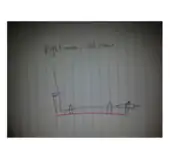 Figure 3: technical drawing of device design |
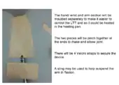 Figure 4: templates for LTT device |
Manufacturing process
Casting method - circumferential cast
- A stockinet was applied over a cutting strip on the patients arms, ensuring to provide enough to cover trim lines
- Important landmarks were marked on the stockinet
- Insure that patient is in the right position
- A 10cm bandage was wrapped around the whole circumference. Starting from the arm moving distally to the hands making sure to overlap one third of the bandage in order to build 3 layers.
- The cast was removed using a plaster saw to cut an opening on the anterior surface along the cutting strip.
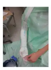 Figure 5: full circumferential cast |
Casting method - back slab
- A stockinet was applied to the patients upper limb and important landmarks and trim lines were marked
- The distance from 10cm proximally to the elbow to metacarpal heads was measured
- Using a 15cm bandage a back slab was made with 4-6 layers
- Ensure patient is in the right position
- Back slab was applied to posterior surface
- As the plaster dried you hold the desired position and mould the plaster
- Removed when dry
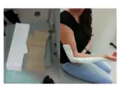 Figure 6 & 7: plaster back slab |
LTT orthotic device
- Templates were used to trace and cut out LTT
- Hand/ wrist section was moulded first
- Soak in warm water until LTT is soft and flexible
- Insure to hold the right position
- LTT was aligned, secured and moulded onto patient
- Trim lines were marked and excess material was cut off
- Next the arm piece was moulded using the same method
- Ensure that the elbow is held in 90 degrees
- Heat up the distal section of the arm piece and the proximal section of the hand/ wrist piece and pinch them together to join.
- Cover the seams with another piece of LTT
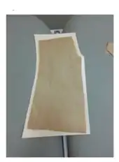 Figure 8: LTT cut outs |
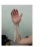 Figure 9: LTT moulded onto patient – hand/ wrist section |
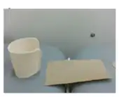 Figure 10: LTT arm section |
Critique of fit
Present client
The patient is a 17-year-old female who has experienced an accident whilst riding her horse. As she fell of her horse she landed on her right hand incorrectly, twisting her wrist. The patient is commencing year 12 VCE and wishes to recover as soon as possible in order to complete her studies efficiently.
Subjective assessment
On presentation, the client reported to experience pain when supinating and pronating her forearm.
Objective assessment
A physical assessment was completed and on observation it was evident that the patient had an excessive prominence of the ulnar head. In assessing movement of the wrist the patient had limited pronation and felt pain with movements associated with the wrist, hence range of motion testing was completed (active, passive and resisted). An x-ray was completed to assess bone integrity and an ultrasound was completed to assess soft tissue dignity.
Diagnosis
Considering the results from the clinical assessment and medical imaging, a likely diagnosis of an isolated dorsal distal radioulnar joint (DRUJ) dislocation (as discussed in Written information – evidence).
Orthotic goals
The goal of orthotic management is to completely immobilise all wrist movements: flexion/ extension and supination/ pronation, and to maintain an optimal healing position: supinated forearm and a neutral wrist.
Prescription
In order to achieve these goals, an orthotic prescription has been developed of an above elbow brace. The device starts proximally to the elbow, runs posteriorly to the arm, and ends distally to the wrist. It is necessary to immobilise the elbow, this will allow control of the forearm and wrist in order to prevent supination and pronation. There is an opening on the anterior surface to allow removal if necessary. There are 4 Velcro straps that suspend the device (shown in figure 3). The elbow is to be held in 90 degrees flexion, the forearm is supinated and the wrist is in neutral, as shown in figure 1 and 2.
Present device
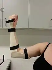 Figure 11: final product (medial view) |
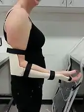 Figure 12: final product (lateral view) |
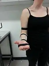 Figure 13: final product (frontal view) |
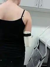 Figure 14: final product (posterior view) |
Has it met prescription design?
- The initial plan was to make an arm piece and a hand wrist piece separately which would then be joined by Velcro straps to suspend the elbow in flexion. This design was unsuccessful, as the arm piece would not stay in position. Therefore a new design was developed.
- As seen in Figure 16 the device does not hold elbow at 90 degrees flexion, in fact the elbow is flexed to 105 degrees. This could have been avoided by ensuring that the patient was in correct position when moulding and joining the two pieces.
- Given the characteristics of LTT, flexion of the elbow and pronation of the DRUJ is still possible. Next time a higher density or stronger material would be used to completely immobilise these movements.
Does it meet high technical standard?
- Joining of the elbow piece and forearm piece looks untidy, figure 14. To prevent this I should’ve designed the elbow piece in a way where it has less seams and so that it would fit to the forearm piece more coherently (elbow piece is square and forearm piece is round).
- The second strap from the proximal end of the device has 2 pieces of Velcro hook. This was a mistake as a result from the initial design.
Outcome measures
Which measures selected, why?
- Using the Disabilities of the arm, shoulder and hand (The DASH) outcome measure survey, relevant questions were chosen that were important for the patient’s everyday activities. Given that the patient has an isolated dorsal distal radioulnar dislocation pronation and supination movements would be affected. Therefore measures that involved pronation and supination movements of the wrist and forearm were included.
- Active range of motion of the wrist (flexion/ extension, pronation/supination) was also assessed and measured.
- Measures would be recorded before the brace was prescribed and again 6-8 weeks after (when the brace is no longer needed)
Did they measure what you had hoped?
- The brace was prescribed to treat the dislocations, therefore it was expected that final outcome measures would have improved from initial outcome measures.
- Yes, the patient’s results improved (shown below in the table 1 and 2)
Were they easily implemented and repeatable?
- These tests were easily implemented as a survey and the survey can be reused.
Reliability
- Surveys are reliable as they measure the patient’s confidence to preform an activity. This self-reporting method allows an easy comparison between performances (Gabel, Michener, Burkett, & Neller, 2006).
TABLE 1: Do you have any difficulties with preforming these tasks using your right (dominant) hand?
1. No difficulty 2. Mild difficulty 3. Moderate difficulty 4. Severe difficulty 5. Unable
| Measure | Initial | Final |
|---|---|---|
| Questions 2 – write | 4 | 1 |
| Question 5 – push open a heavy door | 2 | 1 |
| Question 13 – wash hair | 3 | 1 |
| Question 16 – use a knife to cut food | 4 | 1 |
| Total Score | 13 | 4 |
(Questions were extracted from the Disabilities of the Arm, Shoulder and Hand (DASH) survey)
TABLE 2: Range of motion
| Measure | Initial left | Initial right | Final left |
|---|---|---|---|
| Wrist flexion | 75° | 60° | 70° |
| Wrist extension | 70° | 60° | 65° |
| Pronation | 70° | 40° | 65° |
| Supination | 85° | 80° | 85° |
The outcome measure results shows an restoration of range of motion and improvement in tasks allocated. Range of motion results have improved from the initial results, however they hare not equivalent to the left. This may be due to scar tissue from healing of the TFCC. Results from the selected tasks have also improved, as the patient can complete these tasks with no difficulty. The outcome measure results suggests that the TFCC tear has healed and the DRUJ is now stable.
REFERENCES
Gabel, C. P., Michener, L. A., Burkett, B., & Neller, A. (2006). The upper limb functional index: Development and determination of reliability, validity, and responsiveness. Journal of Hand Therapy, 19(3), 328-48; quiz 349. Retrieved from http://0-search.proquest.com.alpha2.latrobe.edu.au/docview/222239040?accountid=1200
Referral
Addressed to: Stevie Haystack - Physiotherpist, PurePhysio, Level 4, 52 Collins St, Melbourne, CBD, 3000
Re: Patricia Hooves
27 Cobden Avenue, Parahan
To Stevie Haystack,
Thank you for accepting my referral for Miss Hooves. In working together I believe we can achieve optimal results in improving clinical outcome measures and restoring ROM.
Miss Hooves is a 17 y.o who was in an accident whilst riding her horse 2/7 ago. Pt presented with an excessive prominence of the ulnar head and induced pain and loss of ROM with wrist movements of her right hand. X-rays show a dorsally displaced ulnar head but no fractures were present. An ultrasound was also completed, which identified a partial tear of the triangular fibrocartilaginous complex. Pt has been diagnosed with an isolated dorsal distal radioulnar joint dislocation and is currently not on any medication.
I have prescribed and fabricated Miss Hooves a custom above elbow brace for 6 weeks. This device is to be worn at all times, except for when completing rehabilitation exercises. The aim of the device is to immobilise the distal radioulnar joint by also immobilising the elbow and wrist. The brace holds the pt in a stable position, 90 degrees of elbow flexion and forearm supination, which is ideal for correct and effective healing of the injured cartilage.
With your help we hope to restore her full range of motion of the wrist as soon as possible.
I am looking forward to work with you in treating Miss Hooves and in future collaborations.
Kindest regards,
Ms Stephanie Vu
Orthotists, Orthotic specialist centre, 311 High St, Prahran VIC 3181