POLYZOA, in zoology, a term (introduced by J. V. Thompson, 1830) synonymous with Bryozoa (Ehrenberg, 1831) for a group commonly included with the Brachiopoda in the Molluscoidea (Milne Edwards, 1843). The correctness of this association is questionable, and the Polyzoa are here treated as a primary division or phylum of the animal kingdom. They may be defined as aquatic animals, forming colonies by budding; with ciliated retractile tentacles and a U-shaped alimentary canal. The phylum is subdivided as follows.
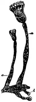
|
| (After van Beneden.) |
| Fig. 1.—Part of the creeping stolon, with zooids, of Pedicellina belgica. a, c, Stalks of zooids of different ages; b, bud. |
Class I. Entoprocta (Nitsche). Lophophore circular, including both mouth and anus. Tentacles infolded, during retraction, into a vestibule which can be closed by a sphincter. Body-wall not calcified, body-cavity absent. Definite excretory organs present. Reproductive organs with ducts leading to the vestibule. Zooids possessing a high degree of individuality. Loxosoma, Pedicellina (fig. 1), Urnatella.
Class II. Ectoprocta (Nitsche). Lophophore circular or horseshoe shaped, including the mouth but not the anus. Tentacles retractile into an introvert (“tentacle-sheath”). Body-wall membranous or calcified, body-cavity distinct. Specific excretory organs absent, with the doubtful exception of the Phylactolaemata. Reproductive organs not continuous with ducts. Zooids usually connected laterally with their neighbours.
Order 1. Gymnolaemata (Allman).—Lophophore circular, with no epistome. Body-cavities of zooids not continuous with one another. Body-wall not muscular.
Sub-order 1. Trepostomata (Ulrich); Fossil.—Zooecia, long and coherent, prismatic or cylindrical, with terminal orifices, their wall thin and simple in structure proximally, thickened and complicated distally. Cavity of the zooecium subdivided by transverse diaphragms, most numerous in the distal portion. Orifices of the zooecia often separated by pores (mesopores).
Sub-order 2. Cryptostomata (Vine); Fossil.—Zooecia usually short. Orifice concealed at the bottom of a vestibular shaft, surrounded by a solid or vesicular calcareous deposit.
Sub-order 3. Cyclostomata (Busk).—Zooecia prismatic or cylindrical, with terminal, typically circular orifice, not protected by any special organ. The ovicells are modified zooecia, and contain numerous embryos which in the cases so far investigated arise by fission of a primary embryo developed from an egg. Crisia (fig. 2), Tubulipora, Hornera, Lichenopora.
Sub-order 4. Ctenostomata (Busk).—Zooecia with soft uncalcified[1] walls, the external part of the introvert being closed during retraction by a membranous collar. Zooecia either arising from a stolon, without lateral connexion with one another, or laterally united to form sheets. Alcyonidium, Flustrella, Bowerbankia (fig. 3), Farrella, Victorella, Paludicella.

|
| (After Hincks.) |
| Fig. 2.—Part of a Branch of Crisia eburnea. g, zooecia; x, imperfectly developed ovicell. |
Sub-order 5. Cheilostomata (Busk).—Zooecia with more or less calcified walls. Orifice closed by a lid-like operculum. Polymorphism usually occurs, certain individuals having the form of avicularia or vibracula. The ovicells commonly found as globular swellings surmounting the orifices are not direct modifications of zooecia, and each typically contains a single egg or embryo. Membranipora, Flustra, Onychocella, Lunulites, Steganoporella, Scrupocellaria, Menipea, Caberea, Bicellaria, Bugula, Beania, Membraniporella, Cribrilina, Cellaria, Micropora, Selenaria, Umbonula (fig. 4), Lepralia, Schizoporella, Cellepora, Mucronella, Smittia, Retepora, Catenicella, Microporella, Adeona.

|
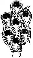
|
| (After Hincks.) | (After Hincks.) |
| Fig. 3.—Part of a branch of Bowerbankia pustulosa, showing the thread-like stolon from which arise young and mature zooecia. The tentacles are expanded in some of the latter. | Fig. 4.—Zooecia of Umbonula pavonella, showing a pair of minute avicularia on either side of the orifice of each zooecium. |
Order 2. Phylactolaemata (Allman).—Lophophore horse-shoe shaped, or in Fredericella circular. Mouth guarded by an epistome. Body-cavities of zooids continuous with one another. Body-wall uncalcified and muscular. Reproduction sexual and by means of “statoblasts,” peculiar internal buds protected by a chitinous shell. Fredericella, Plumatella (fig. 5), Lophopus, Cristatella, Pectinatella.
Hatschek (1888) treated the Entoprocta as a division of his group Scolecida, characterized by the possession of a primary body-cavity and of protonephridia; while he placed the Ectoprocta, with the Phoronida and Brachiopoda, in a distinct group, the Tentaculata. Against this view may be urged the essential similarity between the processes of budding in Entoprocta and Ectoprocta (cf. Seeliger, Zeitschr. wiss. Zool. xlix. 168; l., 560), and the resemblances in the development of the two classes.
Of the forms above indicated there is no palaeontological evidence with regard to the Entoprocta. The Trepostomata are in the main Palaeozoic, although Heteropora, of which recent species exist, is placed by Gregory in this division. The Cryptostomata are also Palaeozoic, and include the abundant and widely-distributed genus Fenestella. The Cyclostomata are numerous in Palaeozoic rocks, but attained a specially predominant position, in the Cretaceous strata, where they are represented by a profusion of genera and species; while they still survive in considerable numbers at the present day. The Ctenostomata are ill adapted for preservation as fossils, though remains referred to this group have been described from Palaeozoic strata. They constitute a small proportion of the recent Polyzoa. The Cheilostomata are usually believed to have made their appearance in the Jurassic period. They are the dominant group at the present day, and are represented by a large number of genera and species. The Phylactolaemata are a small group confined to fresh water, and possess clear indications of adaptation to that habitat. The fresh-water fauna also contains a representative of the Entoprocta (Urnatella), two or three Ctenostomes, such as Victorella and Paludicella, and one or two species of Cheilostomata. With these exceptions, the existing Polyzoa are marine forms, occurring from between tide-marks to abyssal depths in the ocean.

| ||||||||
| (After Allman.) | ||||||||
|
Fig. 5.—Zooid of Plumatella, with expanded tentacles. | ||||||||
|
The Polyzoa are colonial animals, the colony (zoarium) originating in most cases from a free-swimming larva, which attaches itself to some solid object and becomes metamorphosed into the primary individual, or “ancestrula.” In the Phylactolaemata, however, a new colony may originate not only from a larva, but also from a peculiar form of bud known as a statoblast, or by the fission of a fully-developed colony. The ancestrula inaugurates a process of budding, continued by its progeny, and thus gives rise to the mature colony. In Loxosoma the buds break off as soon as they become mature, and a colonial form is thus hardly assumed. In other Entoprocta the buds retain a high degree of individuality, a thread-like stolon giving off the cylindrical stalks, each of which dilates at its end into the body of a zooid. In some of the Ctenostomata the colony is similarly constituted, a branched stolon giving off the zooids, which are not connected with one another. In the majority of Ectoprocta there is no stolon, the zooids growing out of one another and being usually apposed so as to form continuous sheets or branches. In the encrusting type, which is found in a large proportion of the genera, the zooids are usually in a single layer, with their orifices facing away from the substratum; but in certain species the colony becomes multilaminar by the continued superposition of new zooids over the free surfaces of the older ones, whose orifices they naturally occlude. The zoarium may rise up into erect growths composed of a single layer of zooids, the orifices of which are all on one surface, or of two layers of zooids placed back to back, with the orifices on both sides of the fronds or plates. The rigid Cheilostomes which have this habit were formerly placed in the genus Eschara, but the bilaminar type is common to a number of genera, and there can he no doubt that it is not in itself an indication of affinity. The body-wall is extensively calcified in the Cyclostomata and in most Cheilostomata, which may form elegant network-like colonies, as in the unilaminar genus Retepora, or may consist of wavy anastomosing plates, as in the bilaminar Lepralia foliacea of the British coasts, specimens of which may have a diameter of many inches. In other Cheilostomes the amount of calcification may be much less, the supporting skeleton being largely composed of the organic material chitin. In Flustra and other forms belonging to this type, the zoarium is accordingly flexible, and either bilaminar or unilaminar. In many calcareous forms, both Cheilostomes and Cyclostomes, the zoarium is rendered flexible by the interposition of chitinous joints at intervals. This habit is characteristic of the genera Crisia, Cellaria, Catenicella and others, while it occurs in certain species of other genera. The form of the colony may thus be a good generic character, or, on the contrary, a single genus or even species may assume a variety of different forms. While nearly all Polyzoa are permanently fixed to one spot, the colonies of Cristatella and Lophopus among the Phylactolaemata can crawl slowly from place to place.
Anatomy.—The zooids of which the colonies of Ectoprocta are composed consist of two parts: the body-wall and the visceral mass (figs. 6, 9). These were at one time believed to represent two individuals of different kinds, together constituting a zooid. The visceral mass was accordingly, termed the “polypide” and the body-wall which contains it the “zooecium.” This view depended principally on the fact that the life of the polypide and of the zooecium are not coextensive. It is one of the most remarkable facts in the natural history of the Polyzoa that a single zooecium may be tenanted by several polypides, which successively degenerate. The periodical histolysis may be partly due to the absence of specific excretory organs and to the accumulation of pigmented excretory substances in the wall of the alimentary canal. On the degeneration of the polypide, its nutritive material is apparently absorbed for the benefit of the zooid, while the pigmented substances assume a spheroidal form, which either remains as an inert “brown body” in the body-cavity or is discharged to the exterior by the alimentary canal of the new polypide. This is formed as a two-layered “polypide-bud,” which usually develops from the inner side of the zooecial wall, and soon occupies the place of the previous polypide. The inner layer of the polypide-bud gives rise to the structures usually regarded as ectodermic and endodermic, the outer layer to the mesodermic organs.
The polypide consists of a “lophophore” bearing a series of ciliated tentacles by which Diatoms and other microscopic bodies are collected as food, of a U-shaped alimentary canal, and of a central nervous system. While the mouth is invariably encircled by the bases of the tentacles, the anus lies within the series in the Entoprocta and outside it in the Ectoprocta. The lophophore is a simple circle in all Polyzoa except in the Phylactolaemata, where it typically has the form of a horse shoe outlined by the bases of the tentacles. In Fredericella belonging to this order it is, however, circular, but the systematic position of the genus is sufficiently indicated by its possession of an “epistome,” a lip-like structure guarding the anal side of the mouth in all Phylactolaemata and absent throughout the Gymnolaemata. The cavities of the hollow tentacles open into a circular canal which surrounds the oesophagus at the base of the lophophore. This is continuous with the general body-cavity in the Phylactolaemata, while in the Gymnolaemata it develops in the bud as a part of the body-cavity, from which it becomes completely separated. In the Entoprocta the tentacles are withdrawn by being infolded into the “vestibule,” a depression of the oral surface which can be closed by a sphincter muscle. In the Ectoprocta they are retractile into an introvert, the “tentacle-sheath” (fig. 9), the external opening of which is the “orifice” of the zooecium. In the Cyclostomata, further distinguished by the cylindrical or prismatic form of their highly calcified zooecia, the orifice is typically circular, without any definite closing organ. In the Cheilostomata it is closed by a chitinous (rarely calcareous) “operculum” (fig. 9, C), while in the Ctenostomata it is guarded by a delicate membrane similar to a piece of paper rolled into a longitudinally creased cylinder. During retraction this “collar” lies concealed in the beginning of the introvert. It becomes visible when the polypide begins to protrude its tentacles, making its appearance through the orifice as a delicate hyaline frill through which the tentacles are pushed.
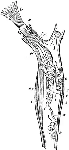
| ||||||||||||||||||||
| (After Allman.) | ||||||||||||||||||||
|
Fig. 6.—Zooid of Paludicella articulata ( = ehrenbergi). | ||||||||||||||||||||
|
In the Phylactolaemata the outermost layer of the body-wall is a flexible uncalcified cuticle or “ectocyst,” beneath which follow in succession the ectoderm, the muscular layers and the coelomic epithelium. In a few Gymnolaemata the ectocyst is merely chitinous, although in most cases the four vertical walls and the basal wall of the zooecium are calcareous. The free (frontal) wall may remain membranous and uncalcified, as in Membranipora (figs. 8 A, 9 A), but in many Cheilostomes the frontal surface is protected by a calcareous shield, which grows from near the free edges of the vertical walls and commonly increases in thickness as the zooecium grows older by the activity of the “epitheca,” a layer of living tissue outside it. The body-wall is greatly simplified in the Gymnolaemata, in correlation with the functional importance of the skeletal part of the wall. Even the ectoderm can rarely be recognized as an obvious epithelium except in regions where budding is taking place, while muscular layers are always absent and a coelomic epithelium can seldom be observed. The body-cavity is, however, traversed by muscles, and by strands of mesodermic “funicular tissue,” usually irregular, but sometimes constituting definite funiculi (fig. 6, x, x′). This tissue is continuous from zooecium to zooecium through perforated “rosette-plates” in the dividing walls. In the Phylactolaemata a single definite funiculus passes from the body-wall to the apex of the stomach. This latter organ is pigmented in all Polyzoa, and is produced, in the Ectoprocta, beyond the point where the intestine leaves it into a conspicuous caecum (fig. 6, v). The nervous system is represented by a ganglion situated between the mouth and the anus. The ovary (o) and the testis (t) of Ectoprocta are developed on the body-wall, on the stomach, or on the funiculus. Both kinds of reproductive organs may occur in a single zooecium, and the reproductive elements pass when ripe into the body-cavity. Their mode of escape is unknown in most cases. In some Gymnolaemata, polypides which develop an ovary possess a flask-shaped “intertentacular organ,” situated between two of the tentacles, and affording a direct passage into the introvert for the eggs or even the spermatozoa developed in the same zooecium. In other cases the reproductive cells perhaps pass out by the atrophy of the polypide, whereby the body-cavity may become continuous with the exterior. The statoblasts of the Phylactolaemata originate on the funiculus, and are said to be derived partly from an ectodermic core possessed by this organ and partly from its external mesoderm (Braem), the former giving rise to the chitinous envelope and to a nucleated layer (fig. 7, ect), which later invaginates to form the inner vesicle of the polypide-bud. The mesodermic portion becomes charged with a yolk-like material (y), and, on the germination of the statoblast, gives rise to the outer layer (mes) of the bud. The production of a polypide by the statoblast thus differs in no essential respect from the formation of a polypide in an ordinary zooecium. The statoblasts require a period of rest before germination, and Braem has shown that their property of floating at the surface may be beneficial to them by exposing them to the action of frost, which in some cases improves the germinating power. The occurrence of Phylactolaemata in the tropics would show, however, without further evidence, that frost is not a factor essential for germination.

| ||||||||||
| (After Braem.) | ||||||||||
|
Fig. 7.—Section of a Germinating Statoblast of Cristatella mucedo. | ||||||||||
|
The withdrawal of the extended polypide is effected by the contraction of the retractor muscles (fig. 6, mr), and must result in an increase in the volume of the contents of the body-cavity. The alternate increase and diminution of volume is easily understood in forms with flexible zooecia. Thus in the Phylactolaemata the contraction of the muscular body-wall exerts a pressure on the fluid of the body-cavity and is the cause of the protrusion of the polypide. In the Gymnolaemata protrusion is effected by the contraction of the parietal muscles, which pass freely across the body-cavity from one part of the body-wall to another. In the branching Ctenostomes the entire body-wall is flexible, so that the contraction of a parietal muscle acts equally on the two points with which it is connected. In encrusting Ctenostomes and in the Membranipora-like Cheilostomes (figs. 8 A, 9 A) the free surface or frontal wall is the only one in which any considerable amount of movement can take place. The parietal muscles (p.m.), which pass from the vertical walls to the frontal wall, thus act by depressing the latter and so exerting a pressure on the fluid of the body-cavity. In Cheilostoma with a rigid frontal wall Jullien showed that protrusion and retraction were rendered possible by the existence of a “compensation-sac,” in communication with the external water.

|
|
Fig. 8.—Diagrammatic Transverse Sections. |
|
A, of Membranipora; B, of an immature zooecium of Cribrilina; p.m., Parietal muscles. |
In its most fully developed condition (fig. 9, C) the compensation-sac (c.s.) is a large cavity which lies beneath the calcified frontal wall and opens to the exterior at the proximal border of the operculum (fig. 10). It is joined to the rigid body-wall by numerous muscle-fibres, the contraction of which must exert a pressure on the fluid of the body-cavity, thereby protruding the polypide. The exchange of fluid in the sac may well have a respiratory significance, in addition to its object of facilitating the movements of the tentacles.

|
|
Fig. 9.—Diagrammatic Longitudinal Sections of Cheilostomatous Zooecia. |
|
A, Membranipora (after Nitsche); B, Cribrilina; C, Some of the Lepralioid forms. b.c., Body-cavity. cr., Cryptocyst. c.s., Compensation-sac. f.m., Frontal membrane. o., Orifice, through which the tentacles are protruded. op., Operculum. p.m., Parietal muscles. t.s., Tentacle-sheath. |
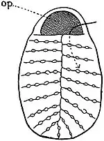
|
|
Fig. 10.—Zooecium of Cribrilina, showing the entrance to the compensation-sac on the proximal side of the operculum (op). |
The evolution of the arrangements for protruding the polypide seems to have proceeded along several distinct lines: (i.) In certain species of Membranipara the “frontal membrane,” or membranous free-wall, is protected by a series of calcareous spines, which start from its periphery and arch inwards. In Cribrilina similar spines are developed in the young zooecium, but they soon unite with one another laterally, leaving rows of pores along the sutural lines (fig. 10). The operculum retains its continuity with the frontal membrane (fig. 9, B) into which the parietal muscles are still inserted. As indications that the conditions described in Membranipora and Cribrilina are of special significance may be noted the fact that the ancestrula of many genera which have well-developed compensation-sacs in the rest of their zooecia is a Membranipora-like individual with a series of marginal calcareous spines, and the further fact that a considerable proportion of the Cretaceous Cheilostomes belong either to the Mernbraniporidae or to the Cribrilinidae. (ii.) In Scrupocellaria, Menipea and Caberea a single, greatly dilated marginal spine, the “scutum” or “fornix,” may protect the frontal membrane. (iii.) In Umbonula the frontal membrane and parietal muscles of the young zooecium are like those of Membranipora, but they become covered by the growth, from the proximal and lateral sides, of a calcareous lamina covered externally by a soft membrane. The arrangement is perhaps derivable from a Cribrilina-like condition in which the outer layer of the spines has become membranous while the spines themselves are laterally united from the first. (iv.) In the Microporidae and Steganoporellidae the body-cavity becomes partially subdivided by a calcareous lamina (“cryptocyst,” Jullien) which grows from the proximal and lateral sides in a plane parallel to the frontal membrane and not far below it. The parietal muscles are usually reduced to a single pair, which may pass through foramina (“opesiules”) in the cryptocyst to reach their insertion. There is no compensation-sac in these families. (v.) Many of the Lepralioid forms offer special difficulties, but the calcareous layer of the frontal surface is probably a cryptocyst (as in fig. 9, C), the compensation-sac being developed round its distal border. The “epitheca” noticed above is in this case the persistent frontal membrane. (vi.) In Microporella the opening of the compensation-sac has become separated from the operculum by calcareous matter, and is known as the “medianipore.” Jullien believed that this pore opens into the tentacle-sheath, but it appears probable that it really communicates with the compensation-sac and not with the tentacle-sheath. The mechanism of protrusion in the Cyclostomata is a subject which requires further examination.
The most singular of the external appendages found in the Polyzoa are the avicularia and vibracula of the Cheilostomata. The avicularium is so called from its resemblance, in its most highly differentiated condition, to the head of a bird. In Bugula, for instance, a calcareous avicularium of this type is attached by a narrow neck to each zooecium. The avicularium can move as a whole by means of special muscles, and its chitinous lower jaw or “mandible” can be opened and closed. It is regarded as a modified zooecium, the polypide of which has become vestigial, although it is commonly represented by a sense-organ, bearing tactile hairs, situated on what may be termed the palate. The operculum of the normal zooecium has become the mandible, while the occlusor muscles have become enormous. In the vibraculum the part representing the zooecium is relatively smaller, and the mandible has become the “seta,” an elongated chitinous lash which projects far beyond the zooecial portion of the structure. In Caberea, the vibracula are known to move synchronously, but co-ordination of this kind is otherwise unknown in the Polyzoa. The avicularia and vibracula give valuable aid to the systematic study of the Cheilostomata. In its least differentiated form the avicularium occupies the place of an ordinary zooecium (“vicarious avicularium”), from which it is distinguished by the greater development of the operculum and its muscles, while the polypide is normally not functional. Avicularia of this type occur in the common Flustra foliacea, in various species of Membranipora, and in particular in the Onychocellidae, a remarkable family common in the Cretaceous period and still existing. In the majority of Cheilostomes, the avicularia are, so to speak, forced out of the ordinary series of zooecia, with which they are rigidly connected. There are comparatively few cases in which, as in Bugula, they are mounted on a movable joint. Although at first sight the arrangement of the avicularia in Cheilostomes appears to follow no general law some method is probably to be made out on closer study. They occur in particular in relation with the orifice of the zooecium, and with that of the compensation-sac. This delicate structure is frequently guarded by an avicularium at its entrance, while avicularia are also commonly found on either side of the operculum or in other positions close to that structure. It can hardly be doubted that the function of these avicularia is the protection of the tentacles and compensation-sac. The suggestion that they are concerned in feeding does not rest on any definite evidence, and is probably erroneous. But avicularia or vibracula may also occur in other places—on the backs of unilaminar erect forms, along the sutural lines of the zooecia and on their frontal surface. These are probably important in checking overgrowth by encrusting organisms, and in particular by preventing larvae from fixing on the zoarium. Vibracula are of less frequent occurrence than avicularia, with which they may coexist as in Scrupocelloria, where they occur on the backs of the unilaminar branches. In the so-called Selenariidae, probably an unnatural association of genera which have assumed a free discoidal form of zoarium, they may reach a very high degree of development, but Busk's suggestion that in this group they “may be subservient to locomotion” needs verification.
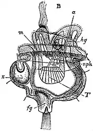
| ||||||||||||||
| (After Hatschek.) | ||||||||||||||
| Fig. 11.—Larva of Pedicellina. | ||||||||||||||
|
Development and Affinities.—It is generally admitted that the larva of the Entoprocta (fig. 11) has the structure of a Trochosphere. This appears to indicate that the Polyzoa are remotely allied to other phyla in which this type of larva prevails, and in particular to the Mollusca and Chaetopoda, as well as to the Rotrfera, which are regarded as persistent Trochospheres. The praeoral portion (lower in fig. 11) constitutes the greater part of the larva and contains most of the viscera. It is terminated by a well-developed structure (fg) corresponding with the apical sense-organ of ordinary Trochospheres, and an excretory organ (nph) of the type familiar in these larvae occurs on the ventral side of the stomach. The central nervous system (x) is highly developed, and in Loxosoma bears a pair of eyes. The larva swims by a ring of cilia, which corresponds with the praeoral circlet of a Trochosphere. The oral surface, on which are situated the mouth (m) an anus (a), is relatively small. The apical sense-organ is used for temporary attachment to the maternal vestibule in which development takes place, but permanent fixation is effected by the oral surface. This is followed by the atrophy of many of the larval organs, including the brain, the sense-organ and the ciliated ring. The alimentary canal persists and revolves in the median plane through an angle of 180°, accompanied by part of the larval vestibule, the space formed by the retraction of the oral surface. The vestibule breaks through to the exterior, and the tentacles, which have been developed within it, are brought into relation with the external water.
In the common and widely-distributed Cheilostome, Membranipora pilosa, the pelagic larva is known as Cyphonautes, and it has a structure not unlike that of the larval Pedicellina. The principal differences are the complication of the ciliated band, the absence of the excretory organ, the great lateral compression of the body, the possession of a pair of shells protecting the sides, the presence of an organ known as the “pyriform organ,” and the occurrence of a sucker in a position corresponding with the depression seen between (m) and (a) in fig. 11. Fixation takes place by means of this sucker, which is everted for the purpose, part of its epithelium becoming the basal ectoderm of the ancestrula. The pyriform organ has probably assisted the larva to find an appropriate place for fixation (cf. Kupelwieser, 18); but, like the alimentary canal and most of the other larval organs, it undergoes a process of histolysis, and the larva becomes the ancestrula, containing the primary brown body derived from the purely larval organs. The polypide is formed, as in an ordinary zooecium after the loss of its polypide, from a polypide-bud.
The Cyphonautes type has been shown by Prouho (24) to occur in two or three widely different species of Cheilostomata and Ctenostomata in which the eggs are laid and develop in the external water. In most Ectoprocta, however, the development takes place internally or in an ovicell, and a considerable quantity of yolk is present. The alimentary canal, which may be represented by a vestigial structure, is accordingly not functional, and the larva does not become pelagic. A pyriform organ is present in most Gymnolaemata as well as the sucker by which fixation is effected. As in the case of Cyphonautes, the larval organs degenerate and the larva becomes the ancestrula from which a polypide is developed as a bud. In the Cyclostomata the primary embryo undergoes repeated fission without developing definite organs, and each of the numerous pieces so formed becomes a free larva, which possesses no alimentary canal. Finally, in the Phylactolaemata, the larva becomes an ancestrula before it is hatched, and one or several polypides may be present when fixation is effected.
The development of the Ectoprocta is intelligible on the hypothesis that the Entoprocta form the starting-point of the series. On the view that the Phylactolaemata are nearly related to Phoronis (see Phoronidea), it is extremely difficult to draw any conclusions with regard to the significance of the facts of development. If the Phylactolaemata were evolved from the type of structure represented by Phoronis or the Pterobranchia (q.v.), the Gymnolaemata should be a further modification of this type, and the comparative study of the embryology of the two orders would appear to be meaningless. It seems more natural to draw the conclusion that the resemblances of the Phylactolaemata to Phoronis are devoid of phylogenetic significance.
Bibliography.—For general accounts of the structure and development of the Polyzoa the reader's attention is specially directed to 12, 14, 6, 25, 1, 2, 17, 26, 18, 23, 3, in the list given below; for an historical account to 1; for a full bibliography of the group, to 22; for fresh-water forms, to 1-3, 7-10, 17; for an indispensable synonymic list of recent marine forms, to 15; for Entoprocta, to 10, 11, 24; for the classification of Gymnolaemata, to 21, 14, 4, 13, 20; for Palaeontology, to 27, 22.
References to important works on the species of marine Polyzoa by Busk, Hincks, Jullien, Levinsen, MacGillivray, Nordgaard, Norman, Waters and others are given in the Memoir (22) by Nickles and Bassler. (1) Allman, “Monogr. Fresh-water Polyzoa,” Ray Soc. (1856). (2) Braem, “Bry. d. süssen Wassers,” Bibl. Zool. Bd. ii. Heft 6 (1890). (3) Braem, “Entwickel. v. Plumatella,” ibid., Bd. x. Heft 23 (1897). (4) Busk, “ Report on the Polyzoa,” “Challenger” Rep. pt. xxx. (1884), 50 (1886). (5) Caldwell, “Phoronis,” Proc. Roy. Soc. (1883), xxxiv. 371. (6) Calvet, “Bry. Ectoproctes Marins,” Trav. Inst. Montpellier (new series), Mém. 8 (1900). (7) Cori, “Nephridien d. Cristatella,” Zeitschr. wiss. Zool. (1893), lv. 626. (8) Davenport, “Cristatella,” Bull. Mus. Harvard (1890-1891), xx. 101. (9) Davenport, “Paludicella,” ibid. (1891-1892), xxii. 1. (10) Davenport, “Urnatella,” ibid. (1893), xxiv. 1. (11) Ehlers, “Pedicellineen,” Abh. Ges. Göttingen (1890), xxxvi. (12) Harmer, “Polyzoa,” Cambr. Nat. Hist. (1896), ii. 463; art. “Polyzoa,” Ency. Brit. (10th ed., 1902), xxxi. 826. (13) Harmer, “Morph. Cheilostomata,” Quart. Journ. Mic. Sci. (1903), xlvi. 263. (14) Hincks, “Hist. Brit. Mar. Pol.” (1880). (15) Jelly, “Syn. Cat. Recent Mar. Bry.” (1889). (16) Jullien and Calvet, “Bryozoaires,” Rés camp. sci. prince de Monaco (1903), xxiii. (17) Kraepelin, “Deutsch. Süsswasser-Bry.,” Abh., Ver. Hamburg (1887), x.; (1892), xii. (18) Kupelwieser, “Cyphonautes,” Zoologica (1906), Bd. xix. Heft 47. (19) Lankester, art. “Polyzoa,” Ency. Brit. (9th ed., 1885), xix. 429. (20) Levinsen, “Bryozoa,” Vid. Medd. Naturh. Foren. (Copenhagen, 1902). (21) MacGillivray, “Cat. Mar. Pol. Victoria,” P. Roy. Soc. Victoria (1887), xxiii, 187. (22) Nickles and Bassler, “Synopsis Amer. Foss. Bry.,” Bull. U.S. Geol. Survey (1900), No. 173. (23) Pace, “Dev. Flustrella,” Quart. Journ. Mic. Soc. (1906), 50, pt. 3, 435. (24) Prouho, “Loxosomes,” Arch. Zool. Exp. (2) (1891), ix. 91. (25) Prouho, “Bryozoaires,” ibid. (2) (1892), x. 557. (26) Seeliger, “Larven u. Verwandtschaft,” Zeitschr. wiss. Zool. (1906), lxxxiv. 1. (27) Ulrich, “Fossil Polyzoa,” in Zittel's Text-book of Palaeontology, Eng. ed. (1900), i. 257.
- (S. F. H.)
- ↑ Calcareous spicules have been described by Lomas in Alcyonidium gelatinosum.