MESOZOA. Van Beneden[1] gave this name to a small group of minute and parasitic animals which he regarded as intermediate between the Protozoa and the Metazoa. The Mesozoa comprise two classes: (1) the Rhombozoa, which are found only in the kidneys of Cephalopods, and (2) the Orthonectida, which infest specimens of Ophiurids, Polychaets, Nemertines, Turbellaria and possibly other groups.

|
|
(From Cambridge Natural History, vol. ii. “Worms, &c.,” by permission of Macmillan & Co. Ltd. After Gamble.) |
|
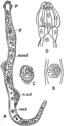
Fig. 1.—Dicyemennea eledones Wag. from the kidney of Eledone moschata. A. Full-grown Rhombogen with infusoriform embryos (emb). g. Part of endoderm cell where formation of the embryos is actively proceeding. n. ect. Nucleus of ectoderm cell. n. end. Nucleus of endoderm cell. p. “Calotte.” B. Developing infusoriform embryo. C. One fully developed. D. “Calotte” of nine cells. |
Class I. Rhombozoa (E. van Beneden).—These animals consist of a central cell from which certain reproductive cells arise, enclosed in a single layer of flattened and for the most part ciliated cells; some of them are modified at the anterior end and form the polar cap. The Rhombozoa comprises two orders: (a) Dicyemida, ciliated vermiform creatures whose polar cap has 8 or 9 cells arranged in two rows (Dicyema, Koll., Dicyemennea, Whitm.); (b) Heterocyemida, non-ciliated animals with no polar cap, but whose anterior ectodermal cells contain refringent bodies and may be produced into wart-like processes (Conuocyema, v. Ben. in Octopus vulgaris; Microcyema in Sepia officinalis). Unlike the Dicyemida, which are fixed in the renal cells of their host by their polar cap, the Heteroqyemida are free. The number of ectoderm cells apart from the polar cap is few, some fourteen to twenty-two.
The central cell is formed by the layer of the first two blastomeres,
and remains quiescent until surrounded by the micromeres or
products of division of the smaller blastomere. It then divides
unequally, and of the two cells thus formed the larger repeats the
process. Each of the two small cells are now called “primary
germ cells,” and they enter into and lie inside the large central
cell. The primary germ cells divide until there are eight of them
all lying within the axial cell. At this stage the future of the
parasite may take one of two directions. Following one path,
the animal (now called a “Nematogen”). gives rise by the segmentation
of its primary germ cells to vermiform larvae which, though
smaller, are but replicas of the parent form. Following the other
path, the animal (now termed a “Rhombogen”), gives origin to a
number of “infusoriform larvae” several of these arising from each
primary germ-cell. The vermiform larvae leave their Nematogen
parent and swimming through the renal fluid attach themselves to
the renal cells. They never leave their host, and die in sea-water.
The infusoriform larvae have a very complicated structure; they
escape from the Rhombogen, and, unlike the vermiform larvae
they can live in sea-water. They possibly serve to infect new
hosts. Some authorities look upon these infusoriform larvae as
males, and consider that they fertilize some of the Nematogens,
which then give rise to males again, whereas the females which
produce the vermiform embryos arise from unfertilized vermiform
larvae. After the infusoriform larvae have left the parent's body,
the Rhombogen takes to producing vermiform offspring, and thus
becomes a secondary Nematogen. Thus, if the above views be
correct, a Rhombogen is a protandrous hermaphrodite.
| ||||||
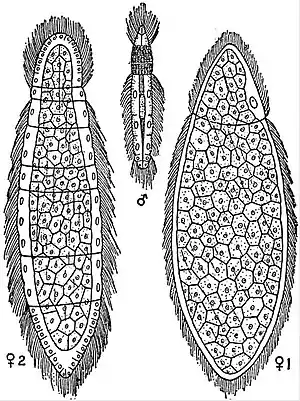
| Fig. 2.—Rhopalura giardii Metschn. from Amphiura squamata. | ||||||
|
E. Nerescheimer has recently described under the name of Lohmanella catenata an organism parasitic in Fritillaria which shows marked affinities, with the Rhombozoa. The genus Haplazoon of which two species have been found in the worms Travisia and Clymene by Dogiel is classed as a new group of Mesozoa.
Class II. Orthonectida (A. Giard).—The Orthonectida contain animals with a central mass of eggs destined to form male and female reproductive cells surrounded by a single layer of ciliated ectoderm cells arranged in regular rings which contain varying numbers of rows of cells. Muscular fibrils occur between the outer and inner cells. The sexes are separate and unlike, and there are two kinds of females, cylindrical and flat. There are but two genera, Rhopalura and Staecharthrum, the latter found in a Polychaet. The male R. giardii lives in the body-cavity of Amphiura squamata, has six rings of ectodermal cells all ciliated except the second, whose cells contain refringent granules. The ectoderm encloses the testis, a mass of cells which have arisen from a single axial cell in the embryo. The female differs from the male in appearance, and in size it is larger. It occurs in two forms: (1) The cylindrical with 8 (or 9) rows of ectoderm cells; here as in the male the second ring is devoid of cilia. (2) The flat females are broader, uniformly ciliated, and have not rings of ectoderm cells. The central mass of cells forms ova which are free, in the cylindrical forms; they leave the mother through the dehiscing of the cells of the non-ciliated ring, are fertilized and develop parthenogenetically into females both flat and cylindrical.
R. spelseneeri and S. giardi are said to be hermaphrodite. The parasites first make their appearance in a host in the form of a plasmodium comparable with the sporocyst of a Trematode. By the segregation of nuclei and some of the surrounding protoplasm, germ cells arise which develop into ciliated larvae an ultimately into males and females which only discharge their spermatozoa and ova when they reach sea-water. The product of the consequent fertilization is unknown; presumably it infects new hosts, entering them in the form of a nucleated plasmodium.
The original idea that in the Rhombozoa and Orthonectida we had animals intermediate between the Protozoa and Metazoa is no longer widely held. The modern view is that the simplicity of their structure is secondary and not primary, and is correlated with their parasitic habit of life. They are probably derived from some Platyhelminthine ancestor and perhaps come nearer to the Trematoda than to any other group.
Literature.—E. van Beneden, Bull. Ac. Belgique (2), (1876) xli. 85, 116; (1876), xlii. 135; also Arch. Biol. (1882), iii. 197; C. O. Whitman, Mt. Stat. Neapel. (188), iv. 1; W. M. Wheeler, Zool. Anz., (1899), xxii. 169; A. Giard, Jour. anat. physiol. (1879), xv. 449; Quart. Jour. Micr. Sci. (1880), xx. 225; St Joseph, Bull. Soc. Zool. France (1896), xxi. 58; Caullery and Mesnil, C. R. ac. sci. (1899), cxxviii. 457 and 516; C. Julin, Arch. Biol. (1882), iii. 1; E. Nerescheimer, Zeitschr. wiss. Zool. (1904), lxxvi. 137; V. A. Dogiel, Trav. soc. imp. natur. St Petersbourg (1907), xxxviii. 28, and Zool. Anz. (1906), xxx. 895.
- (A. E. S.)
- ↑ Bull. Ac. Belgique (1876), p. 35.
