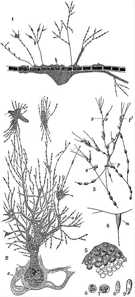
LABYRINTHULIDEA, the name given by Sir Ray Lankester (1885) to Sarcodina (q.v.) forming a reticulate plasmodium, the denser masses united by fine pseudopodical threads, hardly distinct from some Proteomyxa, such as Archerina.
 |
| Labyrinthulidea. |
|
1. A colony or “cell-heap” of Labyrinthula vitellina, Cienk., crawling upon an Alga. 2. A colony or “cell-heap” of Chlamydomyxa labyrinthuloides, Archer, with fully expanded network of threads on which the oat-shaped corpuscles (cells) are moving. o, Is an ingested food particle; at c a portion of the general protoplasm has detached itself and become encysted. 3. A portion of the network of Labyrinthula vitellina, Cienk., more highly magnified. p, Protoplasmic mass apparently produced by fusion of several filaments. p′, Fusion of several cells which have lost their definite spindle-shaped contour. s, Corpuscles which have become spherical and are no longer moving (perhaps about to be encysted). 4. A single spindle cell and threads of Labyrinthula macrocystis, Cienk. n, Nucleus. 5. A group of encysted cells of L. Macrocystis, embedded in a tough secretion. 6, 7. Encysted cells of L. macrocystis, with enclosed protoplasm divided into four spores. 8, 9. Transverse division of a non-encysted spindle-cell of L. macrocystis. |
This is a small and heterogeneous group. Labyrinthula, discovered by L. Cienkowsky, forms a network of relatively stiff threads on which are scattered large spindle-shaped enlargements, each representing an amoeba, with a single nucleus. The threads are pseudopods, very slowly emitted and withdrawn. The amoebae multiply by fission in the active state. The nearest approach to a “reproductive” state is the approximation of the amoebae, and their separate encystment in an irregular heap, recalling the Acrasieae. From each cyst ultimately emerges a single amoeba, or more rarely four (figs. 6, 7). The saprophyte Diplophrys (?) stercorea (Cienk.) appears closely allied to this.
Chlamydomyxa (W. Archer) resembles Labyrinthula in its freely branched plasmodium, but contains yellowish chromatophores, and minute oval vesicles (“physodes”) filled with a substance allied to tannin—possibly phloroglucin—which glide along the plasmodial tracks. The cell-body contains numerous nuclei; but in its active state is not resolvable into distinct oval amoeboids. It is amphitrophic, ingesting and digesting other Protista, as well as “assimilating” by its chromatophores, the product being oil, not starch. The whole body may form a laminated cellulose resting cyst, from which it may only temporarily emerge (fig. 2), or it may undergo resolution into nucleate cells which then encyst, and become multinucleate before rupturing the cyst afresh.
Leydenia (F. Schaudinn) is a parasite in malignant diseases of the pleura. The pseudopodia of adjoining cells unite to form a network; but its affinities seem to such social naked Foraminifera as Mikrogromia.
See Cienkowsky, Archiv f. Microscopische Anatomie, iii. 274 (1867), xii. 44 (1876); W. Archer, Quart. Jour. Microscopic Science, xv. 107 (1875); E. R. Lankester, Ibid., xxxix., 233 (1896); Hieronymus and Jenkinson, Ibid., xiii. 89 (1899); W. Zopf, Beiträge zur Physiologie und Morphologie niederer Organismen, ii. 36 (1892), iv. 60 (1894); Pènard, Archiv für Protistenkunde, iv. 296 (1904); F. Schaudinn and Leyden, Sitzungsberichte der Königlich preussischen Akademie der Wissenschaft, vi. (1896).