GYMNOSPERMS, in Botany. The Gymnosperms, with the Angiosperms, constitute the existing groups of seed-bearing plants or Phanerogams: the importance of the seed as a distinguishing feature in the plant kingdom may be emphasized by the use of the designation Spermophyta for these two groups, in contrast to the Pteridophyta and Bryophyta in which true seeds are unknown. Recent discoveries have, however, established the fact that there existed in the Palaeozoic era fern-like plants which produced true seeds of a highly specialized type; this group, for which Oliver and Scott proposed the term Pteridospermae in 1904, must also be included in the Spermophyta. Another instance of the production of seeds in an extinct plant which further reduces the importance of this character as a distinguishing feature is afforded by the Palaeozoic genus Lepidocarpon described by Scott in 1901; this lycopodiaceous type possessed an integumented megaspore, to which the designation seed may be legitimately applied (see Palaeobotany: Palaeozoic).
As the name Gymnosperm (Gr. γυμνός, naked, σπέρμα, seed) implies, one characteristic of this group is the absence of an ovary or closed chamber containing the ovules. It was the English botanist Robert Brown who first recognized this important distinguishing feature in conifers and cycads in 1825; he established the gymnospermy of these seed-bearing classes as distinct from the angiospermy of the monocotyledons and dicotyledons. As Sachs says in his history of botany, “no more important discovery was ever made in the domain of comparative morphology and systematic botany.” As Coulter and Chamberlain express it, “the habitats of the Gymnosperms to-day indicate that they either are not at home in the more genial conditions affected by Angiosperms, or have not been able to maintain themselves in competition with this group of plants.”
These naked-seeded plants are of special interest on account of their great antiquity, which far exceeds that of the Angiosperms, and as comprising different types which carry us back to the Palaeozoic era and to the forests of the coal period. The best known and by far the largest division of the Gymnosperms is that of the cone-bearing trees (pines, firs, cedars, larches, &c.), which play a prominent part in the vegetation of the present day, especially in the higher latitudes of the northern hemisphere; certain members of this class are of considerable antiquity, but the conifers as a whole are still vigorous and show but little sign of decadence. The division known as the Cycadophyta is represented by a few living genera of limited geographical range and by a large number of extinct types which in the Mesozoic era (see Palaeobotany: Mesozoic) played a conspicuous part in the vegetation of the world. Among existing Cycadophyta we find surviving types which, in their present isolation, their close resemblance to fossil forms, and in certain morphological features, constitute links with the past that not only connect the present with former periods in the earth’s history, but serve as sign-posts pointing the way back along one of the many lines which evolution has followed.
It is needless to discuss at length the origin of the Gymnosperms. The two views which find most favour in regard to the Coniferales and Cycadophyta are: (1) that both have been derived from remote filicinean ancestors; (2) that the cycads are the descendants of a fern-like stock, while conifers have been evolved from lycopodiaceous ancestors. The line of descent of recent cycads is comparatively clear in so far as they have undoubted affinity with Palaeozoic plants which combined cycadean and filicinean features; but opinion is much more divided as to the nature of the phylum from which the conifers are derived. The Cordaitales (see Palaeobotany: Palaeozoic) are represented by extinct forms only, which occupied a prominent position in the Palaeozoic period; these plants exhibit certain features in common with the living Araucarias, and others which invite a comparison with the maidenhair tree (Ginkgo biloba), the solitary survivor of another class of Gymnosperms, the Ginkgoales (see Palaeobotany: Mesozoic). The Gnetales are a class apart, including three living genera, of which we know next to nothing as regards their past history or line of descent. Although there are several morphological features in the three genera of Gnetales which might seem to bring them into line with the Angiosperms, it is usual to regard these resemblances as parallel developments along distinct lines rather than to interpret them as evidence of direct relationship.
Gymnospermae.—Trees or shrubs; leaves vary considerably in size and form. Flowers unisexual, except in a few cases (Gnetales) without a perianth. Monoecious or dioecious. Ovules naked, rarely without carpellary leaves, usually borne on carpophylls, which assume various forms. The single megaspore enclosed in the nucellus is filled with tissue (prothallus) before fertilization, and contains two or more archegonia, consisting usually of a large egg-cell and a small neck, rarely of an egg-cell only and no neck (Gnetum and Welwitschia). Microspore spherical or oval, with or without a bladder-like extension of the exine, containing a prothallus of two or more cells, one of which produces two non-motile or motile male cells. Cotyledons two or several. Secondary xylem and phloem produced by a single cambium, or by successive cambial zones; no true vessels (except in the Gnetales) in the wood, and no companion-cells in the phloem.
| I. | Pteridospermae (see Palaeobotany, Palaeozoic). | |
| II. | Cycadophyta. | |
| A. | Cycadales (recent and extinct). | |
| B. | Bennettitales (see Palaeobotany: Mesozoic). | |
| III. | Cordaitales (see Palaeobotany: Palaeozoic). | |
| IV. | Ginkgoales (recent and extinct). | |
| V. | Coniferales. | |
| A. | Taxaceae. | |
| B. | Pinaceae. | |
There is no doubt that the result of recent research and of work now in progress will be to modify considerably the grouping of the conifers. The family Araucarieae, represented by Araucaria and Agathis, should perhaps be separated as a special class and a rearrangement of other genera more in accord with a natural system of classification will soon be possible; but for the present its twofold subdivision may be retained.
| VI. | Gnetales. | |
| A. | Ephedroideae. | |
| B. | Gnetoideae. | |
| C. | Welwitschioideae (Tumboideae). | |
Cycadophyta.—A. Cycadales.—Stems tuberous or columnar, not infrequently branched, rarely epiphytic (Peruvian species of Zamia); fronds pinnate, bi-pinnate in the Australian genus Bowenia. Dioecious; flowers in the form of cones, except the female flowers of Cycas, which consist of a rosette of leaf-like carpels at the apex of the stem. Seeds albuminous, with one integument; the single embryo, usually bearing two partially fused cotyledons, is attached to a long tangled suspensor. Stems and roots increase in diameter by secondary thickening, the secondary wood being produced by one cambium or developed from successive cambium-rings.
|
|
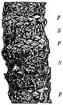
Fig. 1.—Stem of Cycas. F, foliage-leaf bases; S, scale-leaf bases. |
The cycads constitute a homogeneous group of a few living members confined to tropical and sub-tropical regions. As a fairly typical and well-known example of the Cycadaceae, a species of the genus Cycas (e.g. C. circinalis, C. revoluta, &c.) is briefly described. The stout columnar stem may reach a height of 20 metres, and a diameter of half a metre; it remains either unbranched or divides near the summit into several short and thick branches, each branch terminating in a crown of long pinnate leaves. The surface of the stem is covered with rhomboidal areas, which represent the persistent bases of foliage- and scale-leaves. In some species of Cycas there is a well-defined alternation of transverse zones on the stem, consisting of larger areas representing foliage-leaf bases, and similar but smaller areas formed by the bases of scale-leaves (F and S, fig. 1). The scale-leaves clothing the terminal bud are linear-lanceolate in form, and of a brown or yellow colour; they are pushed aside as the stem-axis elongates and becomes shrivelled, finally falling off, leaving projecting bases which are eventually cut off at a still lower level. Similarly, the dead fronds fall off, leaving a ragged petiole, which is afterwards separated from the stem by an absciss-layer a short distance above the base. In some species of Cycas the leaf-bases do not persist as a permanent covering to the stem, but the surface is covered with a wrinkled bark, as in Cycas siamensis, which has a stem of unusual form (fig. 2). Small tuberous shoots, comparable on a large scale with the bulbils of Lycopodium Selago, are occasionally produced in the axils of some of the persistent leaf-bases; these are characteristic of sickly plants, and serve as a means of vegetative reproduction. In the genus Cycas the female flower is peculiar among cycads in consisting of a terminal crown of separate leaf-like carpels several inches in length; the apical portion of each carpellary leaf may be broadly triangular in form, and deeply dissected on the margins into narrow woolly appendages like rudimentary pinnae. From the lower part of a carpel are produced several laterally placed ovules, which become bright red or orange on ripening; the bright fleshy seeds, which in some species are as large as a goose’s egg, and the tawny spreading carpels produce a pleasing combination of colour in the midst of the long dark-green fronds, which curve gracefully upwards and outwards from the summit of the columnar stem. In Cycas the stem apex, after producing a cluster of carpellary leaves, continues to elongate and produces more bud-scales, which are afterwards pushed aside as a fresh crown of fronds is developed. The young leaves of Cycas consist of a straight rachis bearing numerous linear pinnae, traversed by a single midrib; the pinnae are circinately coiled like the leaf of a fern (fig. 3). The male flower of Cycas conforms to the type of structure characteristic of the cycads, and consists of a long cone of numerous sporophylls bearing many oval pollen-sacs on their lower faces. The type described serves as a convenient representative of its class. There are eight other living genera, which may be classified as follows:—
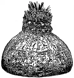
Fig. 2.—Cycas siamensis.
 |
| Fig. 3.—Cycas. Young Frond. |
Classification.—A. Cycadeae.—Characterized by (a) the alternation of scale- and foliage-leaves (fig. 1) on the branched or unbranched stem; (b) the growth of the main stem through the female flower; (c) the presence of a prominent single vein in the linear pinnae; (d) the structure of the female flower, which is peculiar in not having the form of a cone, but consists of numerous independent carpels, each of which bears two or more lateral ovules. Represented by a single genus, Cycas. (Tropical Asia, Australia, &c.).
|
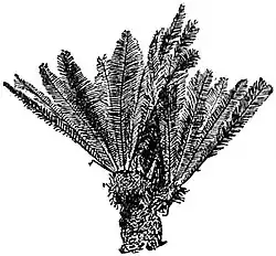
| From a photograph of a plant in Peradeniya Gardens, Ceylon, by Professor R. H. Yapp. |
| Fig. 4.—Dioon edule. |
B. Zamieae.—The stem does not grow through the female flower; both male and female flowers are in the form of cones. (a) Stangerieae.—Characterized by the fern-like venation of the pinnae, which have a prominent midrib, giving off at a wide angle simple or forked and occasionally anastomosing lateral veins. A single genus, Stangeria, confined to South Africa, (b) Euzamieae.—The pinnae are traversed by several parallel veins. Bowenia, an Australian cycad, is peculiar in having bi-pinnate fronds (fig. 5). The various genera are distinguished from one another by the shape and manner of attachment of the pinnae, the form of the carpellary scales, and to some extent by anatomical characters. Encephalartos (South and Tropical Africa).—Large cones; the carpellary scales terminate in a peltate distal expansion. Macrozamia (Australia).—Similar to Encephalartos except in the presence of a spinous projection from the swollen distal end of the carpels. Zamia (South America, Florida, &c.).—Stem short and often divided into several columnar branches. Each carpel terminates in a peltate head. Ceratozamia (Mexico).—Similar in habit to Macrozamia, but distinguished by the presence of two horn-like spinous processes on the apex of the carpels. Microcycas (Cuba).—Like Zamia, except that the ends of the stamens are flat, while the apices of the carpels are peltate. Dioon (Mexico) (fig. 4).—Characterized by the woolly scale-leaves and carpels; the latter terminate in a thick laminar expansion of triangular form, bearing two placental cushions, on which the ovules are situated. Bowenia (Australia).—Bi-pinnate fronds; stem short and tuberous (fig. 5).
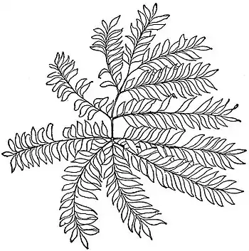
Fig. 5.—Bowenia spectabilis: frond.
|
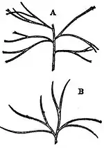
| Fig. 6.—Macrozamia heteromera. A, part of frond; B, single pinna. |
The stems of cycads are often described as unbranched; it is true that in comparison with conifers, in which the numerous branches, springing from the main stem, give a characteristic form to the tree, the tuberous or columnar stem of the Cycadaceae constitutes a striking distinguishing feature. Stem and leaf. Branching, however, occurs not infrequently: in Cycas the tall stem often produces several candelabra-like arms; in Zamia the main axis may break up near the base into several cylindrical branches; in species of Dioon (fig. 4) lateral branches are occasionally produced. The South African Encephalartos frequently produces several branches. Probably the oldest example of this genus in cultivation is in the Botanic Garden of Amsterdam, its age is considered by Professor de Vries to be about two thousand years: although an accurate determination of age is impossible, there is no doubt that many cycads grow very slowly and are remarkable for longevity. The thick armour of petiole-bases enveloping the stem is a characteristic Cycadean feature; in Cycas the alternation of scale-leaves and fronds is more clearly shown than in other cycads; in Encephalartos, Dioon, &c., the persistent scale-leaf bases are almost equal in size to those of the foliage-leaves, and there is no regular alternation of zones such as characterizes some species of Cycas. Another type of stem is illustrated by Stangeria and Zamia, also by a few forms of Cycas (fig. 2), in which the fronds fall off completely, leaving a comparatively smooth stem. The Cyas type of frond, except as regards the presence of a midrib in each pinna, characterizes the cycads generally, except Bowenia and Stangeria. In the monotypic genus Bowenia the large fronds, borne singly on the short and thick stem, are bi-pinnate (fig. 5); the segments, which are broadly ovate or rhomboidal, have several forked spreading veins, and resemble the large pinnules of some species of Adiantum. In Stangeria, also a genus represented by one species (S. paradoxa of South Africa), the long and comparatively broad pinnae, with an entire or irregularly incised margin, are very fern-like, a circumstance which led Kunze to describe the plant in 1835 as a species of the fern Lomaria. In rare cases the pinnae of cycads are lobed or branched: in Dioon spinulosum (Central America) the margin of the segments bears numerous spinous processes; in some species of Encephalartos, e.g. E. horridus, the lamina is deeply lobed; and in a species of the Australian genus Macrozamia, M. heteromera, the narrow pinnae are dichotomously branched almost to the base (fig. 6), and resemble the frond of some species of the fern Schizaea, or the fossil genus Baiera (Ginkgoales). An interesting species of Cycas, C. Micholitzii, has recently been described by Sir William Thiselton-Dyer from Annam, where it was collected by one of Messrs Sanders & Son’s collectors, in which the pinnae instead of being of the usual simple type are dichotomously branched as in Macrozamia heteromera. In Ceratozamia the broad petiole-base is characterized by the presence of two lateral spinous processes, suggesting stipular appendages, comparable, on a reduced scale, with the large stipules of the Marattiaceae among Ferns. The vernation varies in different genera; in Cycas the rachis is straight and the pinnae circinately coiled (fig. 3); in Encephalartos, Dioon, &c., both rachis and segments are straight; in Zamia the rachis is bent or slightly coiled, bearing straight pinnae. The young leaves arise on the stem-apex as conical protuberances with winged borders on which the pinnae appear as rounded humps, usually in basipetal order; the scale-leaves in their young condition resemble fronds, but the lamina remains undeveloped. A feature of interest in connexion with the phylogeny of cycads is the presence of long hairs clothing the scale-leaves, and forming a cap on the summit of the stem-apex or attached to the bases of petioles; on some fossil cycadean plants these outgrowths have the form of scales, and are identical in structure with the ramenta (paleae) of the majority of ferns.
The male flowers of cycads are constructed on a uniform plan, and in all cases consist of an axis bearing crowded, spirally disposed sporophylls. These are often wedge-shaped and angular; in some cases they consist of a short, thick stalk, terminating in a peltate expansion, or prolonged upwards in Flower. the form of a triangular lamina. The sporangia (pollen-sacs), which occur on the under-side of the stamens, are often arranged in more or less definite groups or sori, interspersed with hairs (paraphyses); dehiscence takes place along a line marked out by the occurrence of smaller and thinner-walled cells bounded by larger and thicker-walled elements, which form a fairly prominent cap-like “annulus” near the apex of the sporangium, not unlike the annulus characteristic of the Schizaeaceae among ferns. The sporangial wall, consisting of several layers of cells, encloses a cavity containing numerous oval spores (pollen-grains). In structure a cycadean sporangium recalls those of certain ferns (Marattiaceae, Osmundaceae and Schizaeaceae), but in the development of the spores there are certain peculiarities not met with among the Vascular Cryptogams. With the exception of Cycas, the female flowers are also in the form of cones, bearing numerous carpellary scales. In Cycas revoluta and C. circinalis each leaf-like carpel may produce several laterally attached ovules, but in C. Normanbyana the carpel is shorter and the ovules are reduced to two; this latter type brings us nearer to the carpels of Dioon, in which the flower has the form of a cone, and the distal end of the carpels is longer and more leaf-like than in the other genera of the Zamieae, which are characterized by shorter carpels with thick peltate heads bearing two ovules on the morphologically lower surface. The cones of cycads attain in some cases (e.g. Encephalartos) a considerable size, reaching a length of more than a foot. Cases have been recorded (by Thiselton-Dyer in Encephalartos and by Wieland in Zamia) in which the short carpellary cone-scales exhibit a foliaceous form. It is interesting that no monstrous cycadean cone has been described in which ovuliferous and staminate appendages are borne on the same axis: in the Bennettitales (see Palaeobotany: Mesozoic) flowers were produced bearing on the same axis both androecium and gynoecium.
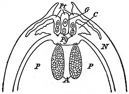 | |
| Fig. 7.—Zamia. Part of Ovule in longitudinal section. (After Webber.) | |
| P, Prothallus. | Pt, Pollen-tube. |
| A, Archegonia. | Pg, Pollen-grain. |
| N, Nucellus. | G,Generative cell (second |
| C, Pollen-chamber. | cell of pollen-tube). |
|
|
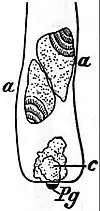
Fig. 8.—Zamia. Proximal end of Pollen-tube, a, a, Spermatozoids from G of fig. 7; Pg, pollen-grain; c, proximal cell (first cell). (After Webber.) |
The pollen-grains when mature consist of three cells, two small and one large cell; the latter grows into the pollen-tube, as in the Coniferales, and from one of the small cells two large ciliated spermatozoids are eventually produced. A remarkable exception to this rule has recently been Microspores and megaspores. recorded by Caldwell, who found that in Microcycas Calocoma the body-cells may be eight or even ten in number and the sperm-cells twice as numerous. One of the most important discoveries made during the latter part of the 19th century was that by Ikeno, a Japanese botanist, who first demonstrated the existence of motile male cells in the genus Cycas. Similar spermatozoids were observed in some species of Zamia by H. J. Webber, and more recent work enables us to assume that all cycads produce ciliated male gametes. Before following the growth of the pollen-grain after pollination, we will briefly describe the structure of a cycadean ovule. An ovule consists of a conical nucellus surrounded by a single integument. At an early stage of development a large cell makes its appearance in the central region of the nucellus; this increases in size and eventually forms three cells; the lowest of these grows vigorously and constitutes the megaspore (embryo-sac), which ultimately absorbs the greater part of the nucellus. The megaspore-nucleus divides repeatedly, and cells are produced from the peripheral region inwards, which eventually fill the spore-cavity with a homogeneous tissue (prothallus); some of the superficial cells at the micropylar end of the megaspore increase in size and divide by a tangential wall into two, an upper cell which gives rise to the short two-celled neck of the archegonium, and a lower cell which develops into a large egg-cell. Each megaspore may contain 2 to 6 archegonia. During the growth of the ovum nourishment is supplied from the contents of the cells immediately surrounding the egg-cell, as in the development of the ovum of Pinus and other conifers. Meanwhile the tissue in the apical region of the nucellus has been undergoing disorganization, which results in the formation of a pollen-chamber (fig. 7, C) immediately above the megaspore. Pollination in cycads has always been described as anemophilous, but according to recent observations by Pearson on South African species it seems probable that, at least in some cases, the pollen is conveyed to the ovules by animal agency. The pollen-grains find their way between the carpophylls, which at the time of pollination are slightly apart owing to the elongation of the internodes of the flower-axis, and pass into the pollen-chamber; the large cell of the pollen-grain grows out into a tube (Pt), which penetrates the nucellar tissue and often branches repeatedly; the pollen-grain itself, with the prothallus-cells, projects freely into the pollen-chamber (fig. 7). The nucleus of the outermost (second) small cell (fig. 7, G) divides, and one of the daughter-nuclei passes out of the cell, and may enter the lowest (first) small cell. The outermost cell, by the division of the remaining nucleus, produces two large spermatozoids (fig. 8, a, a). In Microcycas 16 sperm-cells are produced. In the course of division two bodies appear in the cytoplasm, and behave as centrosomes during the karyokinesis; they gradually become threadlike and coil round each daughter nucleus. This thread gives rise to a spiral ciliated band lying in a depression on the body of each spermatozoid; the large spermatozoids eventually escape from the pollen-tube, and are able to perform ciliary movements in the watery liquid which occurs between the thin papery remnant of nucellar tissue and the archegonial necks. Before fertilization a neck-canal cell is formed by the division of the ovum-nucleus. After the body of a spermatozoid has coalesced with the egg-nucleus the latter divides repeatedly and forms a mass of tissue which grows more vigorously in the lower part of the fertilized ovum, and extends upwards towards the apex of the ovum as a peripheral layer of parenchyma surrounding a central space. By further growth this tissue gives rise to a proembryo, which consists, at the micropylar end, of a sac; the tissue at the chalazal end grows into a long and tangled suspensor, terminating in a mass of cells, which is eventually differentiated into a radicle, plumule and two cotyledons. In the ripe seed the integument assumes the form of a fleshy envelope, succeeded internally by a hard woody shell, internal to which is a thin papery membrane—the apical portion of the nucellus—which is easily dissected out as a conical cap covering the apex of the endosperm. A thorough examination of cycadean seeds has recently been made by Miss Stopes, more particularly with a view to a comparison of their vascular supply with that in Palaeozoic gymnospermous seeds (Flora, 1904). The first leaves borne on the seedling axis are often scale-like, and these are followed by two or more larger laminae, which foreshadow the pinnae of the adult frond.
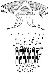
The anatomical structure of the vegetative organs of recent cycads is of special interest as affording important evidence of relationship with extinct types, and with other groups of recent plants. Brongniart, who was the first to investigate in detail the anatomy of a cycadean stem, recognized an agreement, as Anatomy. regards the secondary wood, with Dicotyledons and Gymnosperms, rather than with Monocotyledons. He drew attention also to certain structural similarities between Cycas and Ginkgo. The main anatomical features of a cycad stem may be summarized as follows: the centre is occupied by a large parenchymatous pith traversed by numerous secretory canals, and in some genera by cauline vascular bundles (e.g. Encephalartos and Macrozamia). In addition to these cauline strands (confined to the stem and not connected with the leaves), collateral bundles are often met with in the pith, which form the vascular supply of terminal flowers borne at intervals on the apex of the stem. These latter bundles may be seen in sections of old stems to pursue a more or less horizontal course, passing outwards through the main woody cylinder. This lateral course is due to the more vigorous growth of the axillary branch formed near the base of each flower, which is a terminal structure, and, except in the female flower of Cycas, puts a limit to the apical growth of the stem. The vigorous lateral branch therefore continues the line of the main axis. The pith is encircled by a cylinder of secondary wood, consisting of single or multiple radial rows of tracheids separated by broad medullary rays composed of large parenchymatous cells; the tracheids bear numerous bordered pits on the radial walls. The large medullary rays give to the wood a characteristic parenchymatous or lax appearance, which is in marked contrast to the more compact wood of a conifer. The protoxylem-elements are situated at the extreme inner edge of the secondary wood, and may occur as small groups of narrow, spirally-pitted elements scattered among the parenchyma which abuts on the main mass of wood. Short and reticulately-pitted tracheal cells, similar to tracheids, often occur in the circummedullary region of cycadean stems. In an old stem of Cycas, Encephalartos or Macrozamia the secondary wood consists of several rather unevenly concentric zones, while in some other genera it forms a continuous mass as in conifers and normal dicotyledons. These concentric rings of secondary xylem and phloem (fig. 9) afford a characteristic cycadean feature. After the cambium has been active for some time producing secondary xylem and phloem, the latter consisting of sieve-tubes, phloem-parenchyma and frequently thick-walled fibres, a second cambium is developed in the pericycle; this produces a second vascular zone, which is in turn followed by a third cambium, and so on, until several hollow cylinders are developed. It has been recently shown that several cambium-zones may remain in a state of activity, so that the formation of a new cambium does not necessarily mark a cessation of growth in the more internal meristematic rings. It occasionally happens that groups of xylem and phloem are developed internally to some of the vascular rings; these are characterized by an inverse orientation of the tissues, the xylem being centrifugal and the phloem centripetal in its development. The broad cortical region, which contains many secretory canals, is traversed by numerous vascular bundles (fig. 9, c) some of which pursue a more or less vertical course, and by frequent anastomoses with one another form a loose reticulum of vascular strands; others are leaf-traces on their way from the stele of the stem to the leaves. Most of these cortical bundles are collateral in structure, but in some the xylem and phloem are concentrically arranged; the secondary origin of these bundles from procambium-strands was described by Mettenius in his classical paper of 1860. During the increase in thickness of a cycadean stem successive layers of cork-tissue are formed by phellogens in the persistent bases of leaves (fig. 9, pd), which increase in size to adapt themselves to the growth of the vascular zones. The leaf-traces of cycads are remarkable both on account of their course and their anatomy. In a transverse section of a stem (fig. 9) one sees some vascular bundles following a horizontal or slightly oblique course in the cortex, stretching for a longer or shorter distance in a direction concentric with the woody cylinder. From each leaf-base two main bundles spread right and left through the cortex of the stem (fig. 9, lt), and as they curve gradually towards the vascular ring they present the appearance of two rather flat ogee curves, usually spoken of as the leaf-trace girdles (fig. 9, lt). The distal ends of these girdles give off several branches, which traverse the petiole and rachis as numerous collateral bundles. The complicated girdle-like course is characteristic of the leaf-traces of most recent cycads, but in some cases, e.g. in Zamia floridana, the traces are described by Wieland in his recent monograph on American fossil cycads (Carnegie Institution Publications, 1906) as possessing a more direct course similar to that in Mesozoic genera. A leaf-trace, as it passes through the cortex, has a collateral structure, the protoxylem being situated at the inner edge of the xylem; when it reaches the leaf-base the position of the spiral tracheids is gradually altered, and the endarch arrangement (protoxylem internal) gives place to a mesarch structure (protoxylem more or less central and not on the edge of the xylem strand). In a bundle examined in the basal portion of a leaf the bulk of the xylem is found to be centrifugal in position, but internally to the protoxylem there is a group of centripetal tracheids; higher up in the petiole the xylem is mainly centripetal, the centrifugal wood being represented by a small arc of tracheids external to the protoxylem and separated from it by a few parenchymatous elements. Finally, in the pinnae of the frond the centrifugal xylem may disappear, the protoxylem being now exarch in position and abutting on the phloem. Similarly in the sporophylls of some cycads the bundles are endarch near the base and mesarch near the distal end of the stamen or carpel. The vascular system of cycadean seedlings presents some features worthy of note; centripetal xylem occurs in the cotyledonary bundles associated with transfusion-tracheids. The bundles from the cotyledons pursue a direct course to the stele of the main axis, and do not assume the girdle-form characteristic of the adult plant. This is of interest from the point of view of the comparison of recent cycads with extinct species (Bennettites), in which the leaf-traces follow a much more direct course than in modern cycads. The mesarch structure of the leaf-bundles is met with in a less pronounced form in the flower peduncles of some cycads. This fact is of importance as showing that the type of vascular structure, which characterized the stems of many Palaeozoic genera, has not entirely disappeared from the stems of modern cycads; but the mesarch bundle is now confined to the leaves and peduncles. The roots of some cycads Roots. resemble the stems in producing several cambium-rings; they possess 2 to 8 protoxylem-groups, and are characterized by a broad pericyclic zone. A common phenomenon in cycads is the production of roots which grow upwards (apogeotropic), and appear as coralline branched structures above the level of the ground; some of the cortical cells of these roots are hypertrophied, and contain numerous filaments of blue-green Algae (Nostocaceae), which live as endoparasites in the cell-cavities.
|
|
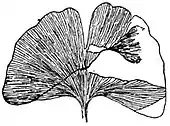
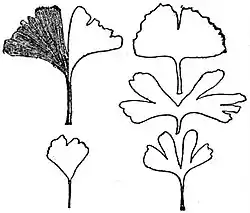
Ginkgoales.—This class-designation has been recently proposed to give emphasis to the isolated position of the genus Ginkgo (Salisburia) among the Gymnosperms. Ginkgo biloba, the maidenhair tree, has usually been placed by botanists in the Taxeae in the neighbourhood of the yew (Taxus), but the proposal by Eichler in 1852 to institute a special family, the Salisburieae, indicated a recognition of the existence of special characteristics which distinguish the genus from other members of the Coniferae. The discovery by the Japanese botanist Hirase of the development of ciliated spermatozoids in the pollen-tube of Ginkgo, in place of the non-motile male cells of typical conifers, served as a cogent argument in favour of separating the genus from the Coniferales and placing it in a class of its own. In 1712 Kaempfer published a drawing of a Japanese tree, which he described under the name Ginkgo; this term was adopted in 1771 by Linnaeus, who spoke of Kaempfer’s plant as Ginkgo biloba. In 1797 Smith proposed to use the name Salisburia adiantifolia in preference to the “uncouth” genus Ginkgo and “incorrect” specific term biloba. Both names are still in common use. On account of the resemblance of the leaves to those of some species of Adiantum, the appellation maidenhair tree has long been given to Ginkgo biloba. Ginkgo is of special interest on account of its isolated position among existing plants, its restricted geographical distribution, and its great antiquity (see Palaeobotany: Mesozoic). This solitary survivor of an ancient stock is almost extinct, but a few old and presumably wild trees are recorded by travellers in parts of China. Ginkgo is common as a sacred tree in the gardens of temples in the Far East, and often cultivated in North America and Europe. Ginkgo biloba, which may reach a height of over 30 metres, forms a tree of pyramidal shape with a smooth grey bark. The leaves (figs. 10 and 11) have a long, slender petiole terminating in a fan-shaped lamina, which may be entire, divided by a median incision into two wedge-shaped lobes, or subdivided into several narrow segments. The venation is like that of many ferns, e.g. Adiantum; the lowest vein in each half of the lamina follows a course parallel to the edge, and gives off numerous branches, which fork repeatedly as they spread in a palmate manner towards the leaf margin. The foliage-leaves occur either scattered on long shoots of unlimited growth, or at the apex of short shoots (spurs), which may eventually elongate into long shoots.

| |
| Fig. 13.—Ginkgo. Apex of Ovule, and Pollen-grain. (After Hirase.) | |
| p, | Pollen-tube (proximal end). |
| c, | Pollen-chamber. |
| e, | Upward prolongation of megaspore. |
| a, | Archegonia. |
| Pg, | Pollen-grain. |
| Ex, | Exine. |
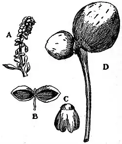
The flowers are dioecious. The male flowers (fig. 12), borne in the axil of scale-leaves, consist of a stalked central axis bearing loosely disposed stamens; each stamen consists of a slender filament terminating in a small apical scale, which bears usually two, but not infrequently three or four pollen-sacs (fig. 12, Flowers. C). The axis of the flower is a shoot bearing leaves in the form of stamens. A mature pollen-grain contains a prothallus of 3 to 5 cells (Fig. 13, Pg); the exine extends over two-thirds of the circumference, leaving a thin portion of the wall, which on collapsing produces a longitudinal groove similar to the median depression on the pollen-grain of a cycad. The ordinary type of female flower has the form of a long, naked peduncle bearing a single ovule on either side of the apex (fig. 12), the base of each being enclosed by a small, collar-like rim, the nature of which has been variously interpreted. A young ovule consists of a conical nucellus surrounded by a single integument terminating as a two-lipped micropyle. A large pollen-chamber occupies the apex of the nucellus; immediately below this, two or more archegonia (fig. 13, a) are developed in the upper region of the megaspore, each consisting of a large egg-cell surmounted by two neck-cells and a canal-cell which is cut off shortly before fertilization. After the entrance of the pollen-grain the pollen-chamber becomes roofed over by a blunt protuberance of nucellar tissue. The megaspore (embryo-sac) continues to grow after pollination until the greater part of the nucellus is gradually destroyed; it also gives rise to a vertical outgrowth, which projects from the apex of the megaspore as a short, thick column (fig. 13, e) supporting the remains of the nucellar tissue which forms the roof of the pollen-chamber (fig. 13, c). Surrounding the pitted wall of the ovum there is a definite layer of large cells, no doubt representing a tapetum, which, as in cycads and conifers, plays an important part in nourishing the growing egg-cell. The endosperm detached from a large Ginkgo ovule after fertilization bears a close resemblance to that of a cycad; the apex is occupied by a depression, on the floor of which two small holes mark the position of the archegonia, and the outgrowth from the megaspore apex projects from the centre as a short peg. After pollination the pollen-tube grows into the nucellar tissue, as in cycads, and the pollen-grain itself (fig. 13, Pg) hangs down into the pollen-chamber; two large spirally ciliated spermatozoids are produced, their manner of development agreeing very closely with that of the corresponding cells in Cycas and Zamia. After fertilization the ovum-nucleus divides and cell-formation proceeds rapidly, especially in the lower part of the ovum, in which the cotyledon and axis of the embryo are differentiated; the long, tangled suspensor of the cycadean embryo is not found in Ginkgo. It is often stated that fertilization occurs after the ovules have fallen, but it has been demonstrated by Hirase that this occurs while the ovules are still attached to the tree. The ripe seed, which grows as large as a rather small plum, is enclosed by a thick, fleshy envelope covering a hard woody shell with two or rarely three longitudinal keels. A papery remnant of nucellus lines the inner face of the woody shell, and, as in cycadean seeds, the apical portion is readily separated as a cap covering the summit of the endosperm.

|
|
Fig. 14.—Ginkgo. Abnormal female Flowers. A, Peduncle; b, scaly bud; B, leaf bearing marginal ovule. (After Fujii.) |
The morphology of the female flowers has been variously interpreted by botanists; the peduncle bearing the ovules has been described as homologous with the petiole of a foliage-leaf and as a shoot-structure, the collar-like envelope at the base of the ovules being referred to as a second integument or arillus, or as the representative of a carpel. The evidence afforded by normal and abnormal flowers appears to be in favour of the following interpretation: The peduncle is a shoot bearing two or more carpels. Each ovule is enclosed at the base by an envelope or collar homologous with the lamina of a leaf; the fleshy and hard coats of the nucellus constitute a single integument. The stalk of an ovule, considerably reduced in normal flowers and much larger in some abnormal flowers, is homologous with a leaf-stalk, with which it agrees in the structure and number of vascular bundles. The facts on which this description is based are derived partly from anatomical evidence, and in part from an account given by a Japanese botanist, Fujii, of several abnormal female flowers; in some cases the collar at the base of an ovule, often described as an arillus, is found to pass gradually into the lamina of a leaf bearing marginal ovules (fig. 14, B). The occurrence of more than two ovules on one peduncle is by no means rare; a particularly striking example is described by Fujii, in which an unusually thick peduncle bearing several stalked ovules terminates in a scaly bud (fig. 14, A, b). The frequent occurrence of more than two pollen-sacs and the equally common occurrence of additional ovules have been regarded by some authors as evidence in favour of the view that ancestral types normally possessed a greater number of these organs than are usually found in the recent species. This Anatomy. view receives support from fossil evidence. Close to the apex of a shoot the vascular bundles of a leaf make their appearance as double strands, and the leaf-traces in the upper part of a shoot have the form of distinct bundles, which in the older part of the shoot form a continuous ring. Each double leaf-trace passes through four internodes before becoming a part of the stele; the double nature of the trace is a characteristic feature. Secretory sacs occur abundantly in the leaf-lamina, where they appear as short lines between the veins; they are abundant also in the cortex and pith of the shoot, in the fleshy integument of the ovule, and elsewhere. The secondary wood of the shoot and root conforms in the main to the coniferous type; in the short shoots the greater breadth of the medullary rays in the more internal part of the xylem recalls the cycadean type. The secondary phloem contains numerous thick-walled fibres, parenchymatous cells, and large sieve-tubes with plates on the radial walls; swollen parenchymatous cells containing crystals are commonly met with in the cortex, pith and medullary-ray tissues. The wood consists of tracheids, with circular bordered pits on their radial walls, and in the late summer wood pits are unusually abundant on the tangential walls. A point of anatomical interest is the occurrence in the vascular bundles of the cotyledons, scale-leaves, and elsewhere of a few centripetally developed tracheids, which give to the xylem-strands a mesarch structure such as characterizes the foliar bundles of cycads. The root is diarch in structure, but additional protoxylem-strands may be present at the base of the main root; the pericycle consists of several layers of cells.
This is not the place to discuss in detail the past history of Ginkgo (see Palaeobotany: Mesozoic). Among Palaeozoic genera there are some which bear a close resemblance to the recent type in the form of the leaves; and petrified Palaeozoic seeds, almost identical with those of the maidenhair tree, have Geological history. been described from French and English localities. During the Triassic and Jurassic periods the genus Baiera—no doubt a representative of the Ginkgoales—was widely spread throughout Europe and in other regions; Ginkgo itself occurs abundantly in Mesozoic and Tertiary rocks, and was a common plant in the Arctic regions as elsewhere during the Jurassic and Lower Cretaceous periods. Some unusually perfect Ginkgo leaves have been found in the Eocene leaf-beds between the lava-flows exposed in the cliffs of Mull (fig. 11). From an evolutionary point of view, it is of interest to note the occurrence of filicinean and cycadean characters in the maidenhair tree. The leaves at once invite a comparison with ferns; the numerous long hairs which form a delicate woolly covering on young leaves recall the hairs of certain ferns, but agree more closely with the long filamentous hairs of recent cycads. The spermatozoids constitute the most striking link with both cycads and ferns. The structure of the seed, the presence of two neck-cells in the archegonia, the late development of the embryo, the partially-fused cotyledons and certain anatomical characters, are features common to Ginkgo and the cycads. The maidenhair tree is one of the most interesting survivals from the past; it represents a type which, in the Palaeozoic era, may have been merged into the extinct class Cordaitales. Through the succeeding ages the Ginkgoales were represented by numerous forms, which gradually became more restricted in their distribution and fewer in number during the Cretaceous and Tertiary periods, terminating at the present day in one solitary survivor.
Coniferales.—Trees and shrubs characterized by a copious branching of the stem and frequently by a regular pyramidal form. Leaves simple, small, linear or short and scale-like, usually persisting for more than one year. Flowers monoecious or dioecious, unisexual, without a perianth, often in the form of cones, but never terminal on the main stem.
The plants usually included in the Coniferae constitute a less homogeneous class than the Cycadaceae. Some authors use the term Coniferae in a restricted sense as including those genera which have the female flowers in the form of cones, the other genera, characterized by flowers of a different External features. type, being placed in the Taxaceae, and often spoken of as Taxads. In order to avoid confusion in the use of the term Coniferae, we may adopt as a class-designation the name Coniferales, including both the Coniferae—using the term in a restricted sense—and the Taxaceae. The most striking characteristic of the majority of the Coniferales is the regular manner of the monopodial branching and the pyramidal shape. Araucaria imbricata, the Monkey-puzzle tree, A. excelsa, the Norfolk Island pine, many pines and firs, cedars and other genera illustrate the pyramidal form. The mammoth redwood tree of California, Sequoia (Wellingtonia) gigantea, which represents the tallest Gymnosperm, is a good example of the regular tapering main stem and narrow pyramidal form. The cypresses afford instances of tall and narrow trees similar in habit to Lombardy poplars. The common cypress (Cupressus sempervirens), as found wild in the mountains of Crete and Cyprus, is characterized by long and spreading branches, which give it a cedar-like habit. A pendulous or weeping habit is assumed by some conifers, e.g. Picea excelsa var. virgata represents a form in which the main branches attain a considerable horizontal extension, and trail themselves like snakes along the ground. Certain species of Pinus, the yews (Taxus) and some other genera grow as bushes, which in place of a main mast-like stem possess several repeatedly-branched leading shoots. The unfavourable conditions in Arctic regions have produced a dwarf form, in which the main shoots grow close to the ground. Artificially induced dwarfed plants of Pinus, Cupressus, Sciadopitys (umbrella pine) and other genera are commonly cultivated by the Japanese. The dying off of older branches and the vigorous growth of shoots nearer the apex of the stem produce a form of tree illustrated by the stone pine of the Mediterranean region (Pinus Pinea), which Turner has rendered familiar in his “Childe Harold’s Pilgrimage” and other pictures of Italian scenery. Conifers are not infrequently seen in which a lateral branch has bent sharply upwards to take the place of the injured main trunk. An upward tendency of all the main lateral branches, known as fastigiation, is common in some species, producing well-marked varieties, e.g. Cephalotaxus pedunculata var. fastigiata; this fastigiate habit may arise as a sport on a tree with spreading branches. Another departure from the normal is that in which the juvenile or seedling form of shoot persists in the adult tree; the numerous coniferous plants known as species of Retinospora are examples of this. The name Retinospora, therefore, does not stand for a true genus, but denotes persistent young forms of Juniperus, Thuja, Cupressus, &c., in which the small scaly leaves of ordinary species are replaced by the slender, needle-like leaves, which stand out more or less at right angles from the branches. The flat branchlets of Cupressus, Thuja (arbor vitae), Thujopsis dolabrata (Japanese arbor vitae) are characteristic of certain types of conifers; in some cases the horizontal extension of the branches induces a dorsiventral structure. A characteristic feature of the genus Agathis (Dammara) the Kauri pine of New Zealand, is the deciduous habit of the branches; these become detached from the main trunk leaving a well-defined absciss-surface, which appears as a depressed circular scar on the stem. A new genus of conifers, Taiwania, has recently been described from the island of Formosa; it is said to agree in habit with the Japanese Cryptomeria, but the cones appear to have a structure which distinguishes them from those of any other genus.
With a few exceptions conifers are evergreen, and retain the leaves for several years (10 years in Araucaria imbricata, 8 to 10 in Picea excelsa, 5 in Taxus baccata; in Pinus the needles usually fall in October of their third year). The larch (Larix) Leaves. sheds its leaves in the autumn, in the Chinese larch (Pseudolarix Kaempferi) the leaves turn a bright yellow colour before falling. In the swamp cypress (Taxodium distichum) the tree assumes a rich brown colour in the autumn, and sheds its leaves together with the branchlets which bear them; deciduous branches occur also in some other species, e.g. Sequoia sempervirens (redwood), Thuja occidentalis, &c. The leaves of conifers are characterized by their small size, e.g. the needle-form represented by Pinus, Cedrus, Larix, &c., the linear flat or angular leaves, appressed to the branches, of Thuja, Cupressus, Libocedrus, &c. The flat and comparatively broad leaves of Araucaria imbricata, A. Bidwillii, and some species of the southern genus Podocarpus are traversed by several parallel veins, as are also the still larger leaves of Agathis, which may reach a length of several inches. In addition to the foliage-leaves several genera also possess scale-leaves of various kinds, represented by bud-scales in Pinus, Picea, &c., which frequently persist for a time at the base of a young shoot which has pushed its way through the yielding cap of protecting scales, while in some conifers the bud-scales adhere together, and after being torn near the base are carried up by the growing axis as a thin brown cap. The cypresses, araucarias and some other genera have no true bud-scales; in some species, e.g. Araucaria Bidwillii, the occurrence of small foliage-leaves, which have functioned as bud-scales, at intervals on the shoots affords a measure of seasonal growth. The occurrence of long and short shoots is a characteristic feature of many conifers. In Pinus the needles occur in pairs, or in clusters of 3 or 5 at the apex of a small and inconspicuous short shoot of limited growth (spur), which is enclosed at its base by a few scale-leaves, and borne on a branch of unlimited growth in the axil of a scale-leaf. In the Californian Pinus monophylla each spur bears usually one needle, but two are not uncommon; it would seem that rudiments of two needles are always produced, but, as a rule, only one develops into a needle. In Sciadopitys similar spurs occur, each bearing a single needle, which in its grooved surface and in the possession of a double vascular bundle bears traces of an origin from two needle-leaves. A peculiarity of these leaves is the inverse orientation of the vascular tissue; each of the two veins has its phloem next the upper and the xylem towards the lower surface of the leaf; this unusual position of the xylem and phloem may be explained by regarding the needle of Sciadopitys as being composed of a pair of leaves borne on a short axillary shoot and fused by their margins (fig. 15, A). Long and short shoots occur also in Cedrus and Larix, but in these genera the spurs are longer and stouter, and are not shed with the leaves; this kind of short shoot, by accelerated apical growth, often passes into the condition of a long shoot on which the leaves are scattered and separated by comparatively long internodes, instead of being crowded into tufts such as are borne on the ends of the spurs. In the genus Phyllocladus (New Zealand, &c.) there are no green foliage-leaves, but in their place flattened branches (phylloclades) borne in the axils of small scale-leaves. The cotyledons are often two in number, but sometimes (e.g. Pinus) as many as fifteen; these leaves are usually succeeded by foliage-leaves in the form of delicate spreading needles, and these primordial leaves are followed, sooner or later, by the adult type of leaf, except in Retinosporas, which retain the juvenile foliage. In addition to the first foliage-leaves and the adult type of leaf, there are often produced leaves which are intermediate both in shape and structure between the seedling and adult foliage. Dimorphism or heterophylly is fairly common. One of the best known examples is the Chinese juniper (Juniperus chinensis), in which branches with spinous leaves, longer and more spreading than the ordinary adult leaf, are often found associated with the normal type of branch. In some cases, e.g. Sequoia sempervirens, the fertile branches bear leaves which are less spreading than those on the vegetative shoots. Certain species of the southern hemisphere genus Dacrydium afford particularly striking instances of heterophylly, e.g. D. Kirkii of New Zealand, in which some branches bear small and appressed leaves, while in others the leaves are much longer and more spreading. A well-known fossil conifer from Triassic strata—Voltzia heterophylla—also illustrates a marked dissimilarity in the leaves of the same shoot. The variation in leaf-form and the tendency of leaves to arrange themselves in various ways on different branches of the same plant are features which it is important to bear in mind in the identification of fossil conifers. In this connexion we may note the striking resemblance between some of the New Zealand Alpine Veronicas, e.g. Veronica Hectori, V. cupressoides, &c. (also Polycladus cupressinus, a Composite), and some of the cypresses and other conifers with small appressed leaves. The long linear leaves of some species of Podocarpus, in which the lamina is traversed by a single vein, recall the pinnae of Cycas; the branches of some Dacrydiums and other forms closely resemble those of lycopods; these superficial resemblances, both between different genera of conifers and between conifers and other plants, coupled with the usual occurrence of fossil coniferous twigs without cones attached to them, render the determination of extinct types a very unsatisfactory and frequently an impossible task.
A typical male flower consists of a central axis bearing numerous spirally-arranged sporophylls (stamens), each of which consists of a slender stalk (filament) terminating distally in a more or less prominent knob or triangular scale, and bearing Flowers. two or more pollen-sacs (microsporangia) on its lower surface. The pollen-grains of some genera (e.g. Pinus) are furnished with bladder-like extensions of the outer wall, which serve as aids to wind-dispersal. The stamens of Araucaria and Agathis are peculiar in bearing several long, and narrow free pollen-sacs; these may be compared with the sporangiophores of the horsetails (Equisetum); in Taxus (yew) the filament is attached to the centre of a large circular distal expansion, which bears several pollen-sacs on its under surface. In the conifers proper the female reproductive organs have the form of cones, which may be styled flowers or inflorescences according to different interpretations of their morphology. In the Taxaceae the flowers have a simpler structure. The female flowers of the Abietineae may be taken as representing a common type. A pine cone reaches maturity in two years; a single year suffices for the full development in Larix and several other genera. The axis of the cone bears numerous spirally disposed flat scales (cone-scales), each of which, if examined in a young cone, is found to be double, and to consist of a lower and an upper portion. The latter is a thin flat scale bearing a median ridge or keel (e.g. Abies), on each side of which is situated an inverted ovule, consisting of a nucellus surrounded by a single integument. As the cone grows in size and becomes woody the lower half of the cone-scale, which we may call the carpellary scale, may remain small, and is so far outgrown by the upper half (seminiferous scale) that it is hardly recognizable in the mature cone. In many species of Abies (e.g. Abies pectinata, &c.) the ripe cone differs from those of Pinus, Picea and Cedrus in the large size of the carpellary scales, which project as conspicuous thin appendages beyond the distal margins of the broader and more woody seminiferous scales; the long carpellary scale is a prominent feature also in the cone of the Douglas pine (Pseudotsuga Douglasii). The female flowers (cones) vary considerably in size; the largest are the more or less spherical cones of Araucaria—a single cone of A. imbricata may produce as many as 300 seeds, one seed to each fertile cone-scale—and the long pendent cones, 1 to 2 ft. in length, of the sugar pine of California (Pinus Lambertiana) and other species. Smaller cones, less than an inch long, occur in the larch, Athrotaxis (Tasmania), Fitzroya (Patagonia and Tasmania), &c. In the Taxodieae and Araucarieae the cones are similar in appearance to those of the Abietineae, but they differ in the fact that the scales appear to be single, even in the young condition; each cone-scale in a genus of the Taxodiinae (Sequoia, &c.) bears several seeds, while in the Araucariinae (Araucaria and Agathis) each scale has one seed. The Cupressineae have cones composed of a few scales arranged in alternate whorls; each scale bears two or more seeds, and shows no external sign of being composed of two distinct portions. In the junipers the scales become fleshy as the seeds ripen, and the individual scales fuse together in the form of a berry. The female flowers of the Taxaceae assume another form; in Microcachrys (Tasmania) the reproductive structures are spirally disposed, and form small globular cones made up of red fleshy scales, to each of which is attached a single ovule enclosed by an integument and partially invested by an arillus; in Dacrydium the carpellary leaves are very similar to the foliage leaves—each bears one ovule with two integuments, the outer of which constitutes an arillus. Finally in the yew, as a type of the family Taxeae, the ovules occur singly at the apex of a lateral branch, enclosed when ripe by a conspicuous red or yellow fleshy arillus, which serves as an attraction to animals, and thus aids in the dispersal of the seeds.
 |
| (C and D after Worsdell.) |
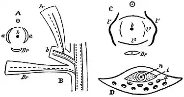
| Fig. 15.—Diagrammatic treatment of:
A, Double needle of Sciadopitys (a, a, leaves; b, shoot; Br, bract). B, seminiferous scale as leaf of axillary shoot (b, shoot; Sc, seminiferous scale; Br, bract). C, seminiferous scale as fused pair of leaves (l1, l2, l3, first, second and third leaves; b, shoot; Br, bract). D, cone-scale of Araucaria (n, nucellus; i, integument; x, xylem). |
It is important to draw attention to some structural features exhibited by certain cone-scales, in which there is no external sign indicative of the presence of a carpellary and a seminiferous scale. In Araucaria Cookii and some allied species each Morphology of female flower. scale has a small pointed projection from its upper face near the distal end; the scales of Cunninghamia (China) are characterized by a somewhat ragged membranous projection extending across the upper face between the seeds and the distal end of the scale; in the scales of Athrotaxis (Tasmania) a prominent rounded ridge occupies a corresponding position. These projections and ridges may be homologous with the seminiferous scale of the pines, firs, cedars, &c. The simplest interpretation of the cone of the Abietineae is that which regards it as a flower consisting of an axis bearing several open carpels, which in the adult cone may be large and prominent or very small, the scale bearing the ovules being regarded as a placental outgrowth from the flat and open carpel. In Araucaria the cone-scale is regarded as consisting of a flat carpel, of which the placenta has not grown out into the scale-like structure. The seminiferous scale of Pinus, &c., is also spoken of sometimes as a ligular outgrowth from the carpellary leaf. Robert Brown was the first to give a clear description of the morphology of the Abietineous cone in which carpels bear naked ovules; he recognized gymnospermy as an important distinguishing feature in conifers as well as in cycads. Another view is to regard the cone as an inflorescence, each carpellary scale being a bract bearing in its axil a shoot the axis of which has not been developed; the seminiferous scale is believed to represent either a single leaf or a fused pair of leaves belonging to the partially suppressed axillary shoot. In 1869 van Tieghem laid stress on anatomical evidence as a key to the morphology of the cone-scales; he drew attention to the fact that the collateral vascular bundles of the seminiferous scale are inversely orientated as compared with those of the carpellary scale; in the latter the xylem of each bundle is next the upper surface, while in the seminiferous scale the phloem occupies that position. The conclusion drawn from this was that the seminiferous scale (fig. 15, B, Sc) is the first and only leaf of an axillary shoot (b) borne on that side of the shoot, the axis of which is suppressed, opposite the subtending bract (fig. 15, A, B, C, Br). Another view is to apply to the seminiferous scale an explanation similar to that suggested by von Mohl in the case of the double needle of Sciadopitys, and to consider the seed-bearing scale as being made up of a pair of leaves (fig. 15, A, a, a) of an axillary shoot (b) fused into one by their posterior margins (fig. 15, A). The latter view receives support from abnormal cones in which carpellary scales subtend axillary shoots, of which the first two leaves (fig. 15, C, l1, l1) are often harder and browner than the others; forms have been described transitional between axillary shoots, in which the leaves are separate, and others in which two of the leaves are more or less completely fused. In a young cone the seminiferous scale appears as a hump of tissue at the base or in the axil of the carpellary scale, but Celakovský, a strong supporter of the axillary-bud theory, attaches little or no importance to this kind of evidence, regarding the present manner of development as being merely an example of a short cut adopted in the course of evolution, and replacing the original production of a branch in the axil of each carpellary scale. Eichler, one of the chief supporters of the simpler view, does not recognize in the inverse orientation of the vascular bundles an argument in support of the axillary-bud theory, but points out that the seminiferous scale, being an outgrowth from the surface of the carpellary scale, would, like outgrowths from an ordinary leaf, naturally have its bundles inversely orientated. In such cone-scales as show little or no external indication of being double in origin, e.g. Araucaria (fig. 15, D) Sequoia, &c., there are always two sets of bundles; the upper set, having the phloem uppermost, as in the seminiferous scale of Abies or Pinus, are regarded as belonging to the outgrowth from the carpellary scale and specially developed to supply the ovules. Monstrous cones are fairly common; these in some instances lend support to the axillary-bud theory, and it has been said that this theory owes its existence to evidence furnished by abnormal cones. It is difficult to estimate the value of abnormalities as evidence bearing on morphological interpretation; the chief danger lies perhaps in attaching undue weight to them, but there is also a risk of minimizing their importance. Monstrosities at least demonstrate possible lines of development, but when the abnormal forms of growth in various directions are fairly evenly balanced, trustworthy deductions become difficult. The occurrence of buds in the axils of carpellary scales may, however, simply mean that buds, which are usually undeveloped in the axils of sporophylls, occasionally afford evidence of their existence. Some monstrous cones lend no support to the axillary-bud theory. In Larix the axis of the cone often continues its growth; similarly in Cephalotaxus the cones are often proliferous. (In rare cases the proliferated portion produces male flowers in the leaf-axils.) In Larix the carpellary scale may become leafy, and the seminiferous scale may disappear. Androgynous cones may be produced, as in the cone of Pinus rigida (fig. 16), in which the lower part bears stamens and the upper portion carpellary and seminiferous scales. An interesting case has been figured by Masters, in which scales of a cone of Cupressus Lawsoniana bear ovules on the upper surface and stamens on the lower face. One argument that has been adduced in support of the axillary bud theory is derived from the Palaeozoic type Cordaites, in which each ovule occurs on an axis borne in the axil of a bract. The whole question is still unsolved, and perhaps insoluble. It may be that the interpretation of the female cone of the Abietineae as an inflorescence, which finds favour with many botanists, cannot be applied to the cones of Agathis and Araucaria. Without expressing any decided opinion as to the morphology of the double cone-scale of the Abietineae, preference may be felt in favour of regarding the cone-scale of the Araucarieae as a simple carpellary leaf bearing a single ovule. A discussion of this question may be found in a paper on the Araucarieae by Seward and Ford, published in the Transactions of the Royal Society of London (1906). Cordaites is an extinct type which in certain respects resembles Ginkgo, cycads and the Araucarieae, but its agreement with true conifers is probably too remote to justify our attributing much weight to the bearing of the morphology of its female flowers on the interpretation of that of the Coniferae. The greater simplicity of the Eichler theory may prejudice us in its favour; but, on the other hand, the arguments advanced in favour of the axillary-bud theories are perhaps not sufficiently cogent to lead us to accept an explanation based chiefly on the uncertain evidence of monstrosities.

|
|
Fig. 16.—Abnormal Cone of Pinus rigida. (After Masters.) |
A pollen-grain when first formed from its mother-cell consists of a single cell; in this condition it may be carried to the nucellus of the ovule (e.g. Taxus, Cupressus, &c.), or more usually (Pinus, Larix, &c.) it reaches maturity before the dehiscence Micro-spores and megaspores. of the microsporangium. The nucleus of the microspore divides and gives rise to a small cell within the large cell, a second small cell is then produced; this is the structure of the ripe pollen-grain in some conifers (Taxus, &c.). The large cell grows out as a pollen-tube; the second of the two small cells (body-cell) wanders into the tube, followed by the nucleus of the first small cell (stalk-cell). In Taxus the body-cell eventually divides into two, in which the products of division are of unequal size, the larger constituting the male generative cell, which fuses with the nucleus of the egg-cell. In Juniperus the products of division of the body-cell are equal, and both function as male generative cells. In the Abietineae cell-formation in the pollen-grain is carried farther. Three small cells occur inside the cavity of the microspore; two of them collapse and the third divides into two, forming a stalk-cell and a larger body-cell. The latter ultimately divides in the apex of the pollen-tube into two non-motile generative cells. Evidence has lately been adduced of the existence of numerous nuclei in the pollen-tubes of the Araucarieae, and it seems probable that in this as in several other respects this family is distinguished from other members of the Coniferales. The precise method of fertilization in the Scots Pine was followed by V. H. Blackman, who also succeeded in showing that the nuclei of the sporophyte generation contain twice as many chromosomes as the nuclei of the gametophyte. Other observers have in recent years demonstrated a similar relation in other genera between the number of chromosomes in the nuclei of the two generations. The ovule is usually surrounded by one integument, which projects beyond the tip of the nucellus as a wide-open lobed funnel, which at the time of pollination folds inwards, and so assists in bringing the pollen-grains on to the nucellus. In some conifers (e.g. Taxus, Cephalotaxus, Dacrydium, &c.) the ordinary integument is partially enclosed by an arillus or second integument. It is held by some botanists (Celakovský) that the seminiferous scale of the Abietineae is homologous with the arillus or second integument of the Taxaceae, but this view is too strained to gain general acceptance. In Araucaria and Saxegothaea the nucellus itself projects beyond the open micropyle and receives the pollen-grains direct. During the growth of the cell which forms the megaspore the greater part of the nucellus is absorbed, except the apical portion, which persists as a cone above the megaspore; the partial disorganization of some of the cells in the centre of the nucellar cone forms an irregular cavity, which may be compared with the larger pollen-chamber of Ginkgo and the cycads. In each ovule one megaspore comes to maturity, but, exceptionally, two may be present (e.g. Pinus sylvestris). It has been shown by Lawson that in Sequoia sempervirens (Annals of Botany, 1904) and by other workers in the genera that several megaspores may attain a fairly large size in one prothallus. The megaspore becomes filled with tissue (prothallus), and from some of the superficial cells archegonia are produced, usually three to five in number, but in rare cases ten to twenty or even sixty may be present. In the genus Sequoia there may be as many as sixty archegonia (Arnoldi and Lawson) in one megaspore; these occur either separately or in some parts of the prothallus they may form groups as in the Cupressineae; they are scattered through the prothallus instead of being confined to the apical region as in the majority of conifers. Similarly in the Araucarieae and in Widdringtonia the archegonia are numerous and scattered and often sunk in the prothallus tissue. In Libocedrus decurrens (Cupressineae) Lawson describes the archegonia as varying in number from 6 to 24 (Annals of Botany xxi., 1907). An archegonium consists of a large oval egg-cell surmounted by a short neck composed of one or more tiers of cells, six to eight cells in each tier. Before fertilization the nucleus of the egg-cell divides and cuts off a ventral canal-cell; this cell may represent a second egg-cell. The egg-cells of the archegonia may be in lateral contact (e.g. Cupressineae) or separated from one another by a few cells of the prothallus, each ovum being immediately surrounded by a layer of cells distinguished by their granular contents and large nuclei. During the development of the egg-cell, food material is transferred from these cells through the pitted wall of the ovum. The tissue at the apex of the megaspore grows slightly above the level of the archegonia, so that the latter come to lie in a shallow depression. In the process of fertilization the two male generative nuclei, accompanied by the pollen-tube nucleus and that of the stalk-cell, pass through an open pit at the apex of the pollen-tube into the protoplasm of the ovum. After fertilization the nucleus of the egg divides, the first stages of karyokinesis being apparent even before complete fusion of the male and female nuclei has occurred. The result of this is the production of four nuclei, which eventually take up a position at the bottom of the ovum and become separated from one another by vertical cell-walls; these nuclei divide again, and finally three tiers of cells are produced, four in each tier. In the Abietineae the cells of the middle tier elongate and push the lowest tier deeper into the endosperm; the cells of the bottom tier may remain in lateral contact and produce together one embryo, or they may separate (Pinus, Juniperus, &c.) and form four potential embryos. The ripe albuminous seed contains a single embryo with two or more cotyledons. The seeds of many conifers are provided with large thin wings, consisting in some genera (e.g. Pinus) of the upper cell-layers of the seminiferous scale, which have become detached and, in some cases, adhere loosely to the seed as a thin membrane; the loose attachment may be of use to the seeds when they are blown against the branches of trees, in enabling them to fall away from the wing and drop to the ground. The seeds of some genera depend on animals for dispersal, the carpellary scale (Microcachrys) or the outer integument being brightly coloured and attractive. In some Abietineae (e.g. Pinus and Picea)—in which the cone-scales persist for some time after the seeds are ripe—the cones hang down and so facilitate the fall of the seeds; in Cedrus, Araucaria and Abies the scales become detached and fall with the seeds, leaving the bare vertical axis of the cone on the tree. In all cases, except some species of Araucaria (sect. Colymbea) the germination is epigean. The seedling plants of some Conifers (e.g. Araucaria imbricata) are characterized by a carrot-shaped hypocotyl, which doubtless serves as a food-reservoir.
The roots of many conifers possess a narrow band of primary xylem-tracheids with a group of narrow spiral protoxylem-elements at each end (diarch). A striking feature in the roots of several genera, excluding the Abietineae, is the occurrence Anatomy. of thick and somewhat irregular bands of thickening on the cell-walls of the cortical layer next to the endodermis. These bands, which may serve to strengthen the central cylinder, have been compared with the netting surrounding the delicate wall of an inflated balloon. It is not always easy to distinguish a root from a stem; in some cases (e.g. Sequoia) the primary tetrarch structure is easily identified in the centre of an old root, but in other cases the primary elements are very difficult to recognize. The sudden termination of the secondary tracheids against the pith-cells may afford evidence of root-structure as distinct from stem-structure, in which the radial rows of secondary tracheids pass into the irregularly-arranged primary elements next the pith. The annual rings in a root are often less clearly marked than in the stem, and the xylem-elements are frequently larger and thinner. The primary vascular bundles in a young conifer stem are collateral, and, like those of a Dicotyledon, they are arranged in a circle round a central pith and enclosed by a common endodermis. It is in the nature of the secondary xylem that the Coniferales are most readily distinguished from the Dicotyledons and Cycadaceae; the wood is homogeneous in structure, consisting almost entirely of tracheids with circular or polygonal bordered pits on the radial walls, more particularly in the late summer wood. In many genera xylem-parenchyma is present, but never in great abundance. A few Dicotyledons, e.g. Drimys (Magnoliaceae) closely resemble conifers in the homogeneous character of the wood, but in most cases the presence of large spring vessels, wood-fibres and abundant parenchyma affords an obvious distinguishing feature.
The abundance of petrified coniferous wood in rocks of various ages has led many botanists to investigate the structure of modern genera with a view to determining how far anatomical characters may be used as evidence of generic distinctions. There are a few well-marked types of wood which serve as convenient standards of comparison, but these cannot be used except in a few cases to distinguish individual genera. The genus Pinus serves as an illustration of wood of a distinct type characterized by the absence of xylem-parenchyma, except such as is associated with the numerous resin-canals that occur abundantly in the wood, cortex and medullary rays; the medullary rays are composed of parenchyma and of horizontal tracheids with irregular ingrowths from their walls. In a radial section of a pine stem each ray is seen to consist in the median part of a few rows of parenchymatous cells with large oval simple pits in their walls, accompanied above and below by horizontal tracheids with bordered pits. The pits in the radial walls of the ordinary xylem-tracheids occur in a single row or in a double row, of which the pits are not in contact, and those of the two rows are placed on the same level. The medullary rays usually consist of a single tier of cells, but in the Pinus type of wood broader medullary rays also occur and are traversed by horizontal resin-canals. In the wood of Cypressus, Cedrus, Abies and several other genera, parenchymatous cells occur in association with the xylem-tracheids and take the place of the resin-canals of other types. In the Araucarian type of wood (Araucaria and Agathis) the bordered pits, which occur in two or three rows on the radial walls of the tracheids, are in mutual contact and polygonal in shape, the pits of the different rows are alternate and not on the same level; in this type of wood the annual rings are often much less distinct than in Cupressus, Pinus and other genera. In Taxus, Torreya (California and the Far East) and Cephalotaxus the absence of resin-canals and the presence of spiral thickening-bands on the tracheids constitute well-marked characteristics. An examination of the wood of branches, stems and roots of the same species or individual usually reveals a fairly wide variation in some of the characters, such as the abundance and size of the medullary rays, the size and arrangement of pits, the presence of wood-parenchyma—characters to which undue importance has often been attached in systematic anatomical work. The phloem consists of sieve-tubes, with pitted areas on the lateral as well as on the inclined terminal walls, phloem-parenchyma and, in some genera, fibres. In the Abietineae the phloem consists of parenchyma and sieve-tubes only, but in most other forms tangential rows of fibres occur in regular alternation with the parenchyma and sieve-tubes. The characteristic companion-cells of Angiosperms are represented by phloem-parenchyma cells with albuminous contents; other parenchymatous elements of the bast contain starch or crystals of calcium oxalate. When tracheids occur in the medullary rays of the xylem these are replaced in the phloem-region by irregular parenchymatous cells known as albuminous cells. Resin-canals, which occur abundantly in the xylem, phloem or cortex, are not found in the wood of the yew. Cephalotaxus (Taxeae) is also peculiar in having resin-canals in the pith (cf. Ginkgo). One form of Cephalotaxus is characterized by the presence of short tracheids in the pith, in shape like ordinary parenchyma, but in the possession of bordered pits and lignified walls agreeing with ordinary xylem-tracheids; it is probable that these short tracheids serve as reservoirs for storing rather than for conducting water. The vascular bundle entering the stem from a leaf with a single vein passes by a more or less direct course into the central cylinder of the stem, and does not assume the girdle-like form characteristic of the cycadean leaf-trace. In species of which the leaves have more than one vein (e.g. Araucaria imbricata, &c.) the leaf-trace leaves the stele of the stem as a single bundle which splits up into several strands in its course through the cortex. In the wood of some conifers, e.g. Araucaria, the leaf-traces persist for a considerable time, perhaps indefinitely, and may be seen in tangential sections of the wood of old stems. The leaf-trace in the Coniferales is simple in its course through the stem, differing in this respect from the double leaf-trace of Ginkgo. A detailed account of the anatomical characters of conifers has been published by Professor D. P. Penhallow of Montreal and Dr. Gothan of Berlin which will be found useful for diagnostic purposes. The characters of leaves most useful for diagnostic purposes are the position of the stomata, the presence and arrangement of resin-canals, the structure of the mesophyll and vascular bundles. The presence of hypodermal fibres is another feature worthy of note, but the occurrence of these elements is too closely connected with external conditions to be of much systematic value. A pine needle grown in continuous light differs from one grown under ordinary conditions in the absence of hypodermal fibres, in the absence of the characteristic infoldings of the mesophyll cell-walls, in the smaller size of the resin-canals, &c. The endodermis in Pinus, Picea and many other genera is usually a well-defined layer of cells enclosing the vascular bundles, and separated from them by a tissue consisting in part of ordinary parenchyma and to some extent of isodiametric tracheids; but this tissue, usually spoken of as the pericycle, is in direct continuity with other stem-tissues as well as the pericycle. The occurrence of short tracheids in close proximity to the veins is a characteristic of coniferous leaves; these elements assume two distinct forms—(1) the short isodiametric tracheids (transfusion-tracheids) closely associated with the veins; (2) longer tracheids extending across the mesophyll at right angles to the veins, and no doubt functioning as representatives of lateral veins. It has been suggested that transfusion-tracheids represent, in part at least, the centripetal xylem, which forms a distinctive feature of cycadean leaf-bundles; these short tracheids form conspicuous groups laterally attached to the veins in Cunninghamia, abundantly represented in a similar position in the leaves of Sequoia, and scattered through the so-called pericycle in Pinus, Picea, &c. It is of interest to note the occurrence of precisely similar elements in the mesophyll of Lepidodendron leaves. An anatomical peculiarity in the veins of Pinus and several other genera is the continuity of the medullary rays, which extend as continuous plates from one end of the leaf to the other. The mesophyll of Pinus and Cedrus is characterized by its homogeneous character and by the presence of infoldings of the cell-walls. In many leaves, e.g. Abies, Tsuga, Larix, &c., the mesophyll is heterogeneous, consisting of palisade and spongy parenchyma. In the leaves of Araucaria imbricata, in which palisade-tissue occurs in both the upper and lower part of the mesophyll, the resin-canals are placed between the veins; in some species of Podocarpus (sect. Nageia) a canal occurs below each vein; in Tsuga, Torreya, Cephalotaxus, Sequoia, &c., a single canal occurs below the midrib; in Larix, Abies, &c., two canals run through the leaf parallel to the margins. The stomata are frequently arranged in rows, their position being marked by two white bands of wax on the leaf-surface.
The chief home of the Coniferales is in the northern hemisphere, where certain species occasionally extend into the Arctic circle and penetrate beyond the northern limit of dicotyledonous trees. Wide areas are often exclusively occupied by Distribution. conifers, which give the landscape a sombre aspect, suggesting a comparison with the forest vegetation of the Coal period. South of the tree-limit a belt of conifers stretches across north Europe, Siberia and Canada. In northern Europe this belt is characterized by such species as Picea excelsa (spruce), which extends south to the mountains of the Mediterranean region; Pinus sylvestris (Scottish fir), reaching from the far north to western Spain, Persia and Asia Minor; Juniperus communis, &c. In north Siberia Pinus Cembra (Cembra or Arolla Pine) has a wide range; also Abies sibirica (Siberian silver fir), Larix sibirica and Juniperus Sabina (savin). In the North American area Picea alba, P. nigra, Larix americana, Abies balsamea (balsam fir), Tsuga canadensis (hemlock spruce), Pinus Strobus (Weymouth pine), Thuja occidentalis (white cedar), Taxus canadensis are characteristic species. In the Mediterranean region occur Cupressus sempervirens, Pinus Pinea (stone pine), species of juniper, Cedrus atlantica, C. Libani, Callitris quadrivalvis, Pinus montana, &c. Several conifers of economic importance are abundant on the Atlantic side of North America—Juniperus virginiana (red cedar, used in the manufacture of lead pencils, and extending as far south as Florida), Taxodium distichum (swamp cypress), Pinus rigida (pitch pine), P. mitis (yellow pine), P. taeda, P. palustris, &c. On the west side of the American continent conifers play a still more striking rôle; among them are Chamaecyparis nutkaensis, Picea sitchensis, Libocedrus decurrens, Pseudotsuga Douglasii (Douglas fir), Sequoia sempervirens, S. gigantea (the only two surviving species of this generic type are now confined to a few localities in California, but were formerly widely spread in Europe and elsewhere), Pinus Coulteri, P. Lambertiana, &c. Farther south, a few representatives of such genera as Abies, Cupressus, Pinus and juniper are found in the Mexican Highlands, tropical America and the West Indies. In the far East conifers are richly represented; among them occur Pinus densiflora, Cryptomeria japonica, Cephalotaxus, species of Abies, Larix, Thujopsis, Sciadopitys verticillata, Pseudolarix Kaempferi, &c. In the Himalaya occur Cedrus deodara, Taxus, species of Cupressus, Pinus excelsa, Abies Webbiana, &c. The continent of Africa is singularly poor in conifers. Cedrus atlantica, a variety of Abies Pinsapo, Juniperus thurifera, Callitris quadrivalvis, occur in the north-west region, which may be regarded as the southern limit of the Mediterranean region. The greater part of Africa north of the equator is without any representatives of the conifers; Juniperus procera flourishes in Somaliland and on the mountains of Abyssinia; a species of Podocarpus occurs on the Cameroon mountains, and P. milanjiana is widely distributed in east tropical Africa. Widdringtonia Whytei, a species closely allied to W. juniperoides of the Cedarberg mountains of Cape Colony, is recorded from Nyassaland and from N.E. Rhodesia; while a third species, W. cupressoides, occurs in Cape Colony. Podocarpus elongata and P. Thunbergii (yellow wood) form the principal timber trees in the belt of forest which stretches from the coast mountains of Cape Colony to the north-east of the Transvaal. Libocedrus tetragona, Fitzroya patagonica, Araucaria brasiliensis, A. imbricata, Saxegothaea and others are met with in the Andes and other regions in South America. Athrotaxis and Microcachrys are characteristic Australian types. Phyllocladus occurs also in New Zealand, and species of Dacrydium, Araucaria, Agathis and Podocarpus are represented in Australia, New Zealand and the Malay regions.
Gnetales.—These are trees or shrubs with simple leaves. The flowers are dioecious, rarely monoecious, provided with one or two perianths. The wood is characterized by the presence of vessels in addition to tracheids. There are no resin-canals. The three existing genera, usually spoken of as members of the Gnetales, differ from one another more than is consistent with their inclusion in a single family; we may therefore better express their diverse characters by regarding them as types of three separate families—(1) Ephedroideae, genus Ephedra; (2) Welwitschioideae, genus Welwitschia; (3) Gnetoideae, genus Gnetum. Our knowledge of the Gnetales leaves much to be desired, but such facts as we possess would seem to indicate that this group is of special importance as foreshadowing, more than any other Gymnosperms, the Angiospermous type. In the more heterogeneous structure of the wood and in the possession of true vessels the Gnetales agree closely with the higher flowering plants. It is of interest to note that the leaves of Gnetum, while typically Dicotyledonous in appearance, possess a Gymnospermous character in the continuous and plate-like medullary rays of their vascular bundles. The presence of a perianth is a feature suggestive of an approach to the floral structure of Angiosperms; the prolongation of the integument furnishes the flowers with a substitute for a stigma and style. The genus Ephedra, with its prothallus and archegonia, which are similar to those of other Gymnosperms, may be safely regarded as the most primitive of the Gnetales. In Welwitschia also the megaspore is filled with prothallus-tissue, but single egg-cells take the place of archegonia. In certain species of Gnetum described by Karsten the megaspore contains a peripheral layer of protoplasm, in which scattered nuclei represent the female reproductive cells; in Gnetum Gnemon a similar state of things exists in the upper half of the megaspore, while the lower half agrees with the megaspore of Welwitschia in being full of prothallus-tissue, which serves merely as a reservoir of food. Lotsy has described the occurrence of special cells at the apex of the prothallus of Gnetum Gnemon, which he regards as imperfect archegonia (fig. 17, C, a); he suggests they may represent vestigial structures pointing back to some ancestral form beyond the limits of the present group. The Gnetales probably had a separate origin from the other Gymnosperms; they carry us nearer to the Angiosperms, but we have as yet no satisfactory evidence that they represent a stage in the direct line of Angiospermic evolution. It is not improbable that the three genera of this ancient phylum survive as types of a blindly-ending branch of the Gymnosperms; but be that as it may, it is in the Gnetales more than in any other Gymnosperms that we find features which help us to obtain a dim prospect of the lines along which the Angiosperms may have been evolved.
Ephedra.—This genus is the only member of the Gnetales represented in Europe. Its species, which are characteristic of warm temperate latitudes, are usually much-branched shrubs. The finer branches are green, and bear a close resemblance to the stems of Equisetum and to the slender twigs of Casuarina; the surface of the long internodes is marked by fine longitudinal ribs, and at the nodes are borne pairs of inconspicuous scale-leaves. The flowers are small, and borne on axillary shoots. A single male flower consists of an axis enclosed at the base by an inconspicuous perianth formed of two concrescent leaves and terminating in two, or as many as eight, shortly stalked or sessile anthers. The female flower is enveloped in a closely fitting sac-like investment, which must be regarded as a perianth; within this is an orthotropous ovule surrounded by a single integument prolonged upwards as a beak-like micropyle. The flower may be described as a bud bearing a pair of leaves which become fused and constitute a perianth, the apex of the shoot forming an ovule. In function the perianth may be compared with a unilocular ovary containing a single ovule; the projecting integument, which at the time of pollination secretes a drop of liquid, serves the same purpose as the style and stigma of an angiosperm. The megaspore is filled with tissue as in typical Gymnosperms, and from some of the superficial cells 3 to 5 archegonia are developed, characterized by long multicellular necks. The archegonia are separated from one another, as in Pinus, by some of the prothallus-tissue, and the cells next the egg-cells (tapetal layer) contribute food-material to their development. After fertilization, some of the uppermost bracts below each flower become red and fleshy; the perianth develops into a woody shell, while the integument remains membranous. In some species of Ephedra, e.g. E. altissima, the fertilized eggs grow into tubular proembryos, from the tip of each of which embryos begin to be developed, but one only comes to maturity. In Ephedra helvetica, as described by Jaccard, no proembryo or suspensor is formed; but the most vigorous fertilized egg, after undergoing several divisions, becomes attached to a tissue, termed the columella, which serves the purpose of a primary suspensor; the columella appears to be formed by the lignification of certain cells in the central region of the embryo-sac. At a later stage some of the cells in the upper (micropylar) end of the embryo divide and undergo considerable elongation, serving the purpose of a secondary suspensor. The secondary wood of Ephedra consists of tracheids, vessels and parenchyma; the vessels are characterized by their wide lumen and by the large simple or slightly-bordered pits on their oblique end-walls.
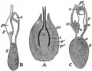
Fig. 17.—Gnetum Gnemon. (After Lotsy.)
|
| |||||||||||||||||||||||||||||
Gnetum.—This genus is represented by several species, most of which are climbing plants, both in tropical America and in warm regions of the Old World. The leaves, which are borne in pairs at the tumid nodes, are oval in form and have a Dicotyledonous type of venation. The male and female inflorescences have the form of simple or paniculate spikes. The spike of an inflorescence bears whorls of flowers at each node in the axils of concrescent bracts accompanied by numerous sterile hairs (paraphyses); in a male inflorescence numerous flowers occur at each node, while in a female inflorescence the number of flowers at each node is much smaller. A male flower consists of a single angular perianth, through the open apex of which the flower-axis projects as a slender column terminating in two anthers. The female flowers, which are more complex in structure, are of two types, complete and incomplete; the latter occur in association with male flowers in a male inflorescence. A complete female flower consists of a nucellus (fig. 17, A, n), surrounded by a single integument (fig. 17, A, i), prolonged upwards as a narrow tube and succeeded by an inner and an outer perianth (fig. 17, A, p′ and p′). The whole flower may be looked upon as an adventitious bud bearing two pairs of leaves; each pair becomes concrescent and forms a perianth, the apex of the shoot being converted into an orthotropous ovule. The incomplete female flowers are characterized by the almost complete suppression of the inner perianth. Several embryo-sacs (megaspores) are present in the nucellus of a young ovule, but one only attains full size, the smaller and partially developed megaspores (fig. 17, B and C, e) being usually found in close association with the surviving and fully-grown megaspore. In Gnetum Gnemon, as described by Lotsy, a mature embryo-sac contains in the upper part a large central vacuole and a peripheral layer of protoplasm, including several nuclei, which take the place of the archegonia of Ephedra; the lower part of the embryo-sac, separated from the upper by a constriction, is full of parenchyma. The upper part of the megaspore may be spoken of as the fertile half (fig. 17, B and C, F) and the lower part, which serves only as food-reservoir for the growing embryo, may be termed the sterile half (fig. 17, B and C, S). (Coulter, Bot. Gazette, xlvi., 1908, regards this tissue as belonging to the nucellus.) At the time of pollination the long tubular integument secretes a drop of fluid at its apex, which holds the pollen-grains, brought by the wind, or possibly to some extent by insect agency, and by evaporation these are drawn on to the top of the nucellus, where partial disorganization of the cells has given rise to an irregular pollen-chamber (fig. 17, A, pc). The pollen-tube, containing two generative and one vegetative nucleus, pierces the wall of the megaspore and then becomes swollen (fig. 17, B and C, pt); finally the two generative nuclei pass out of the tube and fuse with two of the nuclei in the fertile half of the megaspore. As the result of fertilization, the fertilized nuclei of the megaspore become surrounded by a cell-wall, and constitute zygotes, which may attach themselves either to the wall of the megaspore or to the end of a pollen-tube (fig. 17, C, z and z′); they then grow into long tubes or proembryos, which make their way towards the prothallus (C, z′), and eventually embryos are formed from the ends of the proembryo tubes. One embryo only comes to maturity. The embryo of Gnetum forms an out-growth from the hypocotyl, which serves as a feeder and draws nourishment from the prothallus. The fleshy outer portion of the seed is formed from the outer perianth, the woody shell being derived from the inner perianth. The climbing species of Gnetum are characterized by the production of several concentric cylinders of secondary wood and bast, the additional cambium-rings being products of the pericycle, as in Cycas and Macrozamia. The structure of the wood agrees in the main with that of Ephedra.
Welwitschia (Tumboa).—This is by far the most remarkable member of the Gnetales, both as regards habit and the form of its flowers. In a supplement to the systematic work of Engler and Prantl the well-known name Welwitschia, instituted by Hooker in 1864 in honour of Welwitsch, the discoverer of the plant, is superseded by that of Tumboa, originally suggested by Welwitsch. The genus is confined to certain localities in Damaraland and adjoining territory on the west coast of tropical South Africa. A well-grown plant projects less than a foot above the surface of the ground; the stem, which may have a circumference of more than 12 ft., terminates in a depressed crown resembling a circular table with a median groove across the centre and prominent broad ridges concentric with the margin. The thick tuberous stem becomes rapidly narrower, and passes gradually downwards into a tap-root. A pair of small strap-shaped leaves succeed the two cotyledons of the seedling, and persist as the only leaves during the life of the plant; they retain the power of growth in their basal portion, which is sunk in a narrow groove near the edge of the crown, and the tough lamina, 6 ft. in length, becomes split into narrow strap-shaped or thong-like strips which trail on the ground. Numerous circular pits occur on the concentric ridges of the depressed and wrinkled crown, marking the position of former inflorescences borne in the leaf-axil at different stages in the growth of the plant. An inflorescence has the form of a dichotomously-branched cyme bearing small erect cones; those containing the female flowers attain the size of a fir-cone, and are scarlet in colour. Each cone consists of an axis, on which numerous broad and thin bracts are arranged in regular rows; in the axil of each bract occurs a single flower; a male flower is enclosed by two opposite pairs of leaves, forming a perianth surrounding a central sterile ovule encircled by a ring of stamens united below, but free distally as short filaments, each of which terminates in a trilocular anther. The integument of the sterile ovule is prolonged above the nucellus as a spirally-twisted tube expanded at its apex into a flat stigma-like organ. A complete and functional female flower consists of a single ovule with two integuments, the inner of which is prolonged into a narrow tubular micropyle, like that in the flower of Gnetum. The megaspore of Welwitschia is filled with a prothallus-tissue before fertilization, and some of the prothallus-cells function as egg-cells; these grow upwards as long tubes into the apical region of the nucellus, where they come into contact with the pollen-tubes. After the egg-cells have been fertilized by the non-motile male cells they grow into tubular proembryos, producing terminal embryos. The stem is traversed by numerous collateral bundles, which have a limited growth, and are constantly replaced by new bundles developed from strands of secondary meristem. One of the best-known anatomical characteristics of the genus is the occurrence of numerous spindle-shaped or branched fibres with enormously-thickened walls studded with crystals of calcium oxalate. Additional information has been published by Professor Pearson of Cape Town based on material collected in Damaraland in 1904 and 1906–1907. In 1906 he gave an account of the early stages of development of the male and female organs and, among other interesting statements in regard to the general biology of Welwitschia, he expressed the opinion that, as Hooker suspected, the ovules are pollinated by insect-agency. In a later paper Pearson considerably extended our knowledge of the reproduction and gametophyte of this genus.
Authorities.—General: Bentham and Hooker, Genera Plantarum (London, 1862–1883); Engler and Prantl, Die natürlichen Pflanzenfamilien (Leipzig, 1889 and 1897); Strasburger, Die Coniferen und Gnetaceen (Jena, 1872); Die Angiospermen und die Gymnospermen (Jena, 1879); Histologische Beiträge, iv. (Jena, 1892); Coulter and Chamberlain, Morphology of Spermatophytes (New York, 1901); Rendle, The Classification of Flowering Plants, vol. i. (Cambridge, 1904); “The Origin of Gymnosperms” (A discussion at the Linnean Society; New Phytologist, vol. v., 1906). Cycadales: Mettenius, “Beiträge zur Anatomie der Cycadeen,” Abh. k. sächs. Ges. Wiss. (1860); Treub, “Recherches sur les Cycadées,” Ann. Bot. Jard. Buitenzorg, ii. (1884); Solms-Laubach, “Die Sprossfolge der Stangeria, &c.,” Bot. Zeit. xlviii. (1896); Worsdell, “Anatomy of Macrozamia,” Ann. Bot. x. (1896) (also papers by the same author, Ann. Bot., 1898, Trans. Linn. Soc. v., 1900); Scott, “The Anatomical Characters presented by the Peduncle of Cycadaceae,” Ann. Bot. xi. (1897); Lang, “Studies in the Development and Morphology of Cycadean Sporangia, No. I.,” Ann. Bot. xi. (1897); No. II., Ann. Bot. xiv. (1900); Webber, “Development of the Antherozoids of Zamia,” Bot. Gaz. (1897); Ikeno, “Untersuchungen über die Entwickelung, &c., bei Cycas revoluta,” Journ. Coll. Sci. Japan, xii. (1898); Wieland, “American Fossil Cycads,” Carnegie Institution Publication (1906); Stopes, “Beiträge zur Kenntnis der Fortpflanzungsorgane der Cycadeen,” Flora (1904); Caldwell, “Microcycas Calocoma,” Bot. Gaz. xliv., 1907 (also papers on this and other Cycads in the Bot. Gaz., 1907–1909); Matte, Recherches sur l’appareil libéro-ligneux des Cycadacées (Caen, 1904). Ginkgoales: Hirase, “Études sur la fécondation, &c., de Ginkgo biloba,” Journ. Coll. Sci. Japan, xii. (1898); Seward and Gowan, “Ginkgo biloba,” Ann. Bot. xiv. (1900) (with bibliography); Ikeno, “Contribution à l’étude de la fécondation chez le Ginkgo biloba,” Ann. Sci. Nat. xiii. (1901); Sprecher, Le Ginkgo biloba (Geneva, 1907). Coniferales: “Report of the Conifer Conference” (1891) Journ. R. Hort. Soc. xiv. (1892); Beissner, Handbuch der Nadelholzkunde (Berlin, 1891); Masters, “Comparative Morphology of the Coniferae,” Journ. Linn. Soc. xxvii. (1891); ibid. (1896), &c.; Penhallow, “The Generic Characters of the North American Taxaceae and Coniferae,” Proc. and Trans. R. Soc. Canada, ii. (1896); Blackman, “Fertilization in Pinus sylvestris,” Phil. Trans. (1898) (with bibliography); Worsdell, “Structure of the Female Flowers in Conifers,” Ann. Bot. xiv. (1900) (with bibliography); ibid. (1899); Veitch, Manual of the Coniferae (London, 1900); Penhallow, “Anatomy of North American Coniferales,” American Naturalist (1904); Engler and Pilger, Das Pflanzenreich, Taxaceae (1903); Seward and Ford, “The Araucarieae, recent and extinct,” Phil. Trans. R. Soc. (1906) (with bibliography); Lawson, “Sequoia sempervirens,” Annals of Botany (1904); Robertson, “Torreya Californica,” New Phytologist (1904); Coker, “Gametophyte and Embryo of Taxodium,” Bot. Gazette (1903); E. C. Jeffrey, “The Comparative Anatomy and Phylogeny of the Coniferales, part i. The Genus Sequoia,” Mem. Boston Nat. Hist. Soc. v. No. 10 (1903); Gothan, “Zur Anatomie lebender und fossiler Gymnospermen-Hölzer,” K. Preuss. Geol. Landes. (Berlin, 1905) (for more recent papers, see Ann. Bot., New Phytologist, and Bot. Gazette, 1906–1909). Gnetales: Hooker, “On Welwitschia mirabilis.” Trans. Linn. Soc. xxiv. (1864); Bower, “Germination, &c., in Gnetum,” Journ. Mic. Sci. xxii. (1882); ibid. (1881); Jaccard, “Recherches embryologiques sur l’Ephedra helvetica,” Diss. Inaug. Lausanne (1894); Karsten, “Zur Entwickelungsgeschichte der Gattung Gnetum,” Cohn’s Beiträge, vi. (1893); Lotsy, “Contributions to the Life-History of the genus Gnetum,” Ann. Bot. Jard. Buitenzorg, xvi. (1899); Land, “Ephedra trifurca,” Bot. Gazette (1904); Pearson, “Some observations on Welwitschia mirabilis,” Phil. Trans. R. Soc. (1906); Pearson, “Further Observations on Welwitschia,” Phil. Trans. R. Soc. vol. 200 (1909). (A. C. Se.)
