CYSTOFLAGELLATA (so named by E. Haeckel), a group
of Mastigophorous Protozoa, distinguished from Flagellata by
their large size (0.15–1.5 mm.), and their branched endoplasm,
recalling that of Trachelius among Infusoria, within a firm
ectosarc bounded by a strong cuticle. Nutrition is holozoic,
a deep groove leading down to a mouth and pharynx. A long
fine flagellum arises from the pharynx in Noctiluca (E. Suriray)
and Leptodiscus and (R. Hertwig); and in the former genus, a
second flagellum, thick, long and transversely striated, rises
farther out, in the groove; this was likened by E. R. Lankester
to a proboscis, whence his name of Rhynchoflagellata, which
we discard as unnecessary and posterior to Haeckel’s. Noctiluca
has thus the form of an apple with a long stalk. Leptodiscus
(R. Hertwig) has the form of a medusa without a proboscis—it
is menisciform with the thin contractile margin produced
inwards like a velum on the concave side, while the mouth is on
the convex surface and the single flagellum springs from a blind tube on the same surface. Craspedotella (C. A. Kofoid), the
third genus, is still more medusiform, with a broad velum, and
the mouth in a convex central protrusion of the roof of the bell;
and a thick flagellum springs from a blind tube on the convex
surface. All three genera are pelagic and phosphorescent,
this property being seated in the ectoplasm; Noctiluca miliaris
is indeed the chief source of the phosphorescence of our summer
seas. O. Bütschli, like other writers, regards the Cystoflagellates
as closely allied to the Dinoflagellates, the small flagellum corresponding to the longitudinal, the large flagellum to the
transverse flagellum of that group.
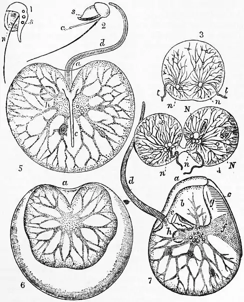
 | |
| After E. Ray Lankester, Ency. Brit., 9th ed. | |
| Cystoflagellate Protozoa. | |
| 1 and 2, Young stages of Noctiluca miliaris.
a, the big flagellum; the unlettered filament becomes the oral flagellum of the adult.
n, nucleus.
s, the so-called spine (superficial ridge of the adult).
3 and 4, Two stages in the fission of Noctiluca miliaris, Suriray. 5. Noctiluca miliaris, viewed from the aboral side (after Allman, Quart. Jour. Mic. Sci., 1872). a, entrance to atrium or flagellar fossa (= longitudinal groove of Dinoflagellata).
c, superficial ridge.
d, big flagellum (= flagellum of transverse groove of Dinoflagellata).
h, nucleus. |
6. Noctiluca miliaris, acted upon by iodine solution, showing the protoplasm shrunk away from the structureless pellicle.
a = entrance to atrium. 7. Lateral view of Noctiluca miliaris. e = mouth and gullet, in which is seen Krohn’s oral flagellum (= the chief flagellum, or flagellum of the longitudinal groove of Dinoflagellata).
f, broad process of protoplasm extending from the superficial ridge c to the central protoplasm.
g, duplicature of pellicle in connexion with superficial ridge.
h, nucleus. |
The reproduction of Noctiluca has been fairly made out; in the adult state it divides by fission down the oral groove; as a preliminary the external differentiations disappear, and the nucleus divides by modified mitosis; then the external organs are regenerated. Under circumstances not well made out, conjugation between two adults takes place by their fusion commencing at the oral region; flagella and pharynx disappear and the nuclei fuse, while the cytoplasts condense into a sphere. The nucleus undergoes broad division, the young nuclei pass to the surface, which becomes imperfectly divided by grooves into as many rounded prominences as there are nuclei (up to 128 or 256); and these become constricted off from the residual useless cytoplasm as zoospores with two unequal flagella, which were at first regarded as Dinoflagellates, of which they have the form (figs. 5, 6). The metamorphosis of these has not yet been observed.
Literature.—E. Suriray, Magazin de zoologie, 1836; G. J. Allman, Quarterly Journal of Microscopic Science, n.s. xii., 1872; L. Cienkowsky, “Zoospore formation in Noctiluca,” Archiv f. mikroskopische Anatomie, vii., 1871; R. Hertwig, “Leptodiscus,” Jenaische Zeitschrift, xi., 1877; C. Ischikawa, Journal of the College of Science (Tokyo, 1894), xii., 1899; F. Doflein, “Conjugation of Noctiluca,” Zoologische Jahrbücher, Anatomie, xiv., 1900; C. A. Kofoid, “Craspedotella,” in Bull. Mus. Comp. Zool. Harvard, xlvi., 1905; O. Bütschli, “Mastigophora,” in Protozoa (Braun’s Thierreich, vol. i., Protozoa) (1883–1887). (M. Ha.)