| |
|
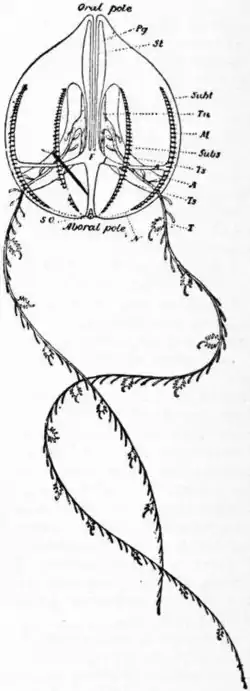
Fig. 1.—Schematic drawing of a Cydippid from the side. (After Chun.) | |
| A, | Adradial canals. |
| F, | Infundibulum. |
| I, | Interradial canal. |
| M, | Meridianal canal lying under a costa. |
| N, | Ciliated furrow from sense pole to costa. |
| Pg, | Paragastric canal. |
| SO, | Sense-organ. |
| St, | Stomodaeum. |
| Subs, | Subsagittal costa. |
| Subt, | Subtentacular costa. |
| T, | Tentacle. |
| Ts, | Boundaries of tentacle-sheath. |
CTENOPHORA, in zoology, a class of jelly-fish which were briefly described by Professor T. H. Huxley in 1875 (see Actinozoa, Ency. Brit. 9th ed. vol. i.) as united with what we now term Anthozoa to form the group Actinozoa; but little was known of the intimate structure of those remarkable and beautiful forms till the appearance in 1880 of C. Chun’s Monograph of the Ctenophora occurring in the Bay of Naples. They may be defined as Coelentera which exhibit both a radial and bilateral symmetry of organs; with a stomodaeum; with a mesenchyma which is partly gelatinous but partly cellular; with eight meridianal rows of vibratile paddles formed of long fused or matted cilia; lacking nematocysts (except in one genus). An example common on the British coasts is furnished by Hormiphora (Cydippe). In outward form this is an egg-shaped ball of clear jelly, having a mouth at the pointed (oral) pole, and a sense-organ at the broader (aboral) pole. It possesses eight meridians (costae) of iridescent paddles in constant vibration, which run from near one pole towards the other; it has also two pendent feathery tentacles of considerable length, which can be retracted into pouches. The mouth leads into an ectodermal stomodaeum (“stomach”), and the latter into an endodermal funnel (infundibulum); these two are compressed in planes at right angles to one another, the sectional long axis of the stomodaeum lying in the so-called sagittal (stomodaeal or gastric) plane, that of the funnel in the transverse (tentacular or funnel) plane. From the funnel, canals are given off in three directions; (a) a pair of paragastric (stomachal, or stomodaeal) canals run orally, parallel to the stomodaeum, and end blindly near the mouth; (b) a pair of perradial canals run in the transverse plane towards the equator of the animal; each of these becomes divided into two short canals at the base of the tentacle sheath which they supply, but has previously given off a pair of short interradial canals, which again bifurcate into two adradial canals; all these branches lie in the equatorial plane of the animal, but the eight adradial canals then open into eight meridianal canals which run orally and aborally under the costae; (c) a pair of aboral vessels which run towards the sense-organ, each of which bifurcates; of the four vessels thus formed, two only open at the sides of the sense-organ, forming the so-called excretory apertures. These three sets of structures, with the funnel from which they rise, make up the endodermal coelenteron, or gastro-vascular system. The generative organs are endodermal by origin, borne at the sides of the meridianal canals as indicated by the signs ♂ ♀. There exists a subepithelial plexus with nerve cells and fibres, similar to that of jelly-fishes. The sense-organ of the aboral pole is complex, and lies under a dome of fused cilia shaped like an inverted bell-jar; it consists of an otolith, formed of numerous calcareous spheroids, which is supported on four plates of fused cilia termed balancers, but is otherwise free. The ciliated ectoderm below the organ is markedly thickened, and perhaps functionally represents a nerve-ganglion: from it eight ciliated furrows radiate outwards, two passing under each balancer as through an archway, and diverge each to the head of a meridianal costa. These ciliated furrows stain deeply with osmic acid, and nervous impulses are certainly transmitted along them. Locomotion is effected by strokes of the paddles in an aboral direction, driving the animal mouth forwards through the water: each paddle or comb (Gr. κτείς; hence Ctenophora) consists of a plate of fused or matted cilia set transversely to the costa. The myoepithelial cells (formerly termed neuro-muscular cells), characteristic of other Coelentera, are not to be found in this group. On the other hand there are well-marked muscle fibres in definite layers, derived from special mesoblastic cells in the embryo, which are embedded in a jelly; these in their origin and arrangement are quite comparable to the mesoderm of Triploblastica, and, although the muscle-cells of some jelly-fish exhibit a somewhat similar condition, nothing so highly specialized as the mesenchyme of Ctenophora occurs in any other Coelenterate. The nematocysts being nearly absent from their group, their chief function is carried out by adhesive lasso-cells.

| |
|
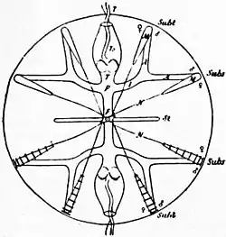
Fig. 2.—Schematic drawing of a Cydippid from the aboral pole. (After Chun.) | |
| T | (centrally), Tentacular canal, and (distally) tentacle. |
| ♂, | Position of testes. |
| ♀, | Position of ovaries; other letters in fig. 1. The stomodaeum lies in the sagittal plane, the funnel and tentacles in the transverse or tentacular plane. |
The Ctenophora are classified as follows:—
| Sub-class i. Tentaculata, | Order | 1. Cydippidea, | Hormiphora. |
| ” | 2. Lobata, | Deiopea. | |
| ” | 3. Cestoidea, | Cestus. | |
| ” ii. Nuda, | ” | Beroë. |
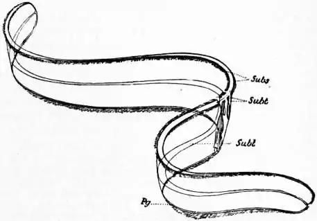
The Tentaculata, as the name implies, may be recognized by the presence of tentacles of some sort. The Cydippidea are generally spherical or ovoid, with two long retrusible pinnate tentacles: the meridianal and paragastric canals end blindly. An example of these has already been briefly described. The Lobata are of the same general type as the first Order, except for the presence of four circumoral auricles (processes of the subtransverse costae) and of a pair of sagittal outgrowths or lobes, on to which the subsagittal costae are continued. Small accessory tentacles lie in grooves, but there is no tentacular pouch; the meridianal vessels anastomose in the lobes. In the Cestoidea the body is compressed in the transverse plane, elongated in the sagittal plane, so as to become riband-like: the subtransverse costae are greatly reduced, the subsagittal costae extend along the aboral edge of the riband. The subsagittal canals lie immediately below their costae aborally, but continuations of the subtransverse canals round down the middle of the riband, and at its end unite, not only with the subsagittal but also with the paragastric canals which run along the oral edge of the riband. The tentacular bases and pouches are present, but there is no main tentacle as in Cydippidea; fine accessory tentacles lie in four grooves along the oral edge. The sub-class Nuda have no tentacles of any kind; they are conical or ovoid, with a capacious stomodaeum like the cavity of a thimble. There is a coelenteric network formed by anastomoses of the meridianal and paragastric canals all over the body.
The embryology of Callianira has been worked out by E. Mechnikov. Segmentation is complete and unequal, producing macromeres and micromeres marked by differences in the size and in yolk-contents. The micromeres give rise to the ectoderm; each of the sixteen macromeres, after budding off a small mesoblast cell, passes on as endoderm. A gastrula is established by a mixed process of embole and epibole. The mesoblast cells travel to the aboral pole of the embryo, and there form a cross-shaped mass, the arms of which lie in the sagittal and transverse planes (perradii).
 | |
| Fig. 3.—Schematic Drawing of Cestus. (After Chun.) | |
| Subs, Subsagittal costae. Subt, Much reduced subtentacular costae. Subt, Branch of the subtentacular canal which runs along the centre of the riband. |
Pg, Continuation of the paragastric canal at right angles to its original direction along the lower edge of the riband. At the right-hand end the last two are seen to unite with the subsagittal canal. |
There can be but little question of the propriety of including Ctenophora among the Coelentera. The undivided coelenteron (gastro-vascular system) which constitutes the sole cavity of the body, the largely radial symmetry, the presence of endodermal generative organs on the coelenteric canals, the subepithelial nerve-plexus, the mesogloea-like matrix of the body—all these features indicate affinity to other Coelentera, but, as has been stated in the article under that title, the relation is by no means close. At what period the Ctenophora branched off from the line of descent, which culminated in the Hydromedusae and Scyphozoa of to-day, is not clear, but it is practically certain that they did so before the point of divergence of these two groups from one another. The peculiar sense-organ, the specialization of the cilia into paddles with the corresponding modifications of the coelenteron, the anatomy and position of the tentacles, and, above all, the character and mode of formation of the mesenchyme, separate them widely from other Coelentera.
 |
| Fig. 4.—Schematic Drawing of Beröe. (After Chun.) |
The last-named character, however, combined with the discovery of two remarkable organisms, Coeloplana and Ctenoplana, has suggested affinity to the flat-worms termed Turbellaria. Ctenoplana, the best known of these, has recently been redescribed by A. Willey (Quart. Journ. Micr. Sci. xxxix., 1896). It is flattened along the axis which unites sense-organ and mouth, so as to give it a dorsal (aboral) surface, and a ventral (oral) surface on which it frequently creeps. Its costae are very short, and retrusible; its two tentacles are pinnate and are also retrusible. Two crescentic rows of ciliated papillae lie in the transverse plane on each side of the sense-organ. The coelenteron exhibits six lobes, two of which Willey identifies with the stomodaeum of other Ctenophora; the other four give rise to a system of anastomosing canals such as are found in Beroë and Polyclad Turbellaria. An aboral vessel embraces the sense-organ, but has no external opening. Ctenoplana is obviously a Ctenophoran flattened and of a creeping habit. Coeloplana is of similar form and habit, with two Ctenophoran tentacles: it has no costae, but is uniformly ciliated. These two forms at least indicate a possible stepping-stone from Ctenophora to Turbellaria, that is to say, from diploblastic to triploblastic Metazoa. By themselves they would present no very weighty argument for this line of descent from two-layered to three-layered forms, but the coincidences which occur in the development of Ctenophora and Turbellaria,—the methods of segmentation and gastrulation, of the separation of the mesoblast cells, and of mesenchyme formation,—together with the marked similarity of the adult mesenchyme in the two groups, have led many to accept this pedigree. In his Monograph on the Polyclad Turbellaria of the Bay of Naples, A. Lang regards a Turbellarian, so to say, as a Ctenophora, in which the sensory pole has rotated forwards in the sagittal plane through 90° as regards the original oral-aboral axis, a rotation which actually occurs in the development of Thysanozoon (Müller’s larva); and he sees, in the eight lappets of the preoral ciliated ring of such a larva, the rudiments of the costal plates. According to his view, a simple early Turbellarian larva, such as that of Stylochus, most nearly represents for us to-day that ancestor from which Ctenophora and Turbellaria are alike derived. For details of this brilliant theory, the reader is referred to the original monograph.
Literature.—G. C. Bourne, “The Ctenophora,” in Ray Lankester’s Treatise on Zoology (1900), where a bibliography is given; G. Curreri, “Osservazioni sui ctenofori,” Boll. Soc. Zool. Ital. (2), i. pp. 190-193 et ii. pp. 58-76; A. Garbe, “Untersuchungen über die Entstehung der Geschlechtsorgane bei den Ctenophoren.,” Zeitschr. Wiss. Zool. lxix. pp. 472-491; K. C. Schneider, Lehrbuch der vergleich. Histologie (1902). (G. H. Fo.)